Medical Imaging of Normal and Pathologic Anatomy (Original PDF from Publisher)
Medical Imaging of Normal and Pathologic Anatomy (Original PDF from Publisher)
| Publisher |
Elsevier |
|---|---|
| Language |
English |
| Edition |
1st |
| Format |
Publisher PDF |
| ISBN-10 |
1437706347 |
| ISBN-13 |
978-1437706345, 9781437706345 |
$33.00 Original price was: $33.00.$22.00Current price is: $22.00.
- The files will be sent to you via E-mail
- Once you placed your order, we will make sure that you receive the files as soon as possible
5/5
Description
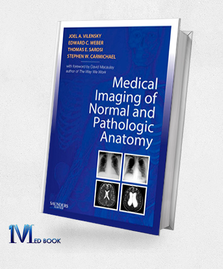
Medical Imaging of Normal and Pathologic Anatomy (Original PDF from Publisher)
Crafted to cater to the contemporary medical student and seamlessly complementing existing gross Medical Imaging of Normal and Pathologic Anatomy, this novel pictorial handbook, authored by Drs. Vilensky, Weber, Carmichael, and Sarosi, delivers an innovative approach to swiftly recognizing pathologic counterparts of gross anatomy.
Medical Imaging of Normal and Pathologic Anatomy presents an extensive array of high-quality radiographic, MR, CT, and ultrasound images in tandem with both normal and pathologic conditions.
This resource expedites the development of proficiency in distinguishing between physiological and abnormal states.
It further offers insights into the selection of suitable imaging modalities for various pathologies, a vital skill for diverse clinical contexts.
In addition, this visual guide markedly enhances performance in academic courses, as well as pivotal assessments such as the USMLE and NBME final exams.
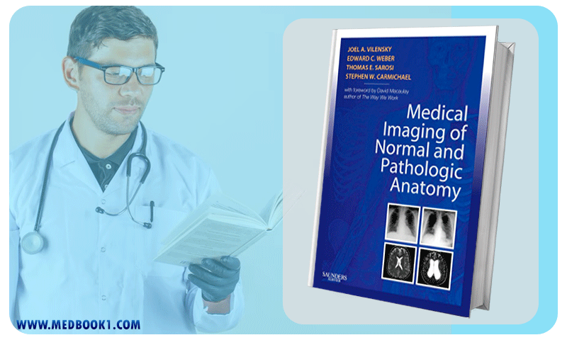
Medical Imaging of Normal and Pathologic Anatomy (Original PDF from Publisher)
Medical Imaging of Normal and Pathologic Anatomy distinctly showcases side-by-side radiography, MR, CT, and ultrasound images, expertly elucidating both normal and anomalous anatomical states.
This adeptly aids in rapid identification of conditions while simultaneously refining diagnostic competencies.
Encompassing clinical conditions commonly featured in core textbooks, the resource adroitly juxtaposes these scenarios with radiologic illustrations of the clinical correlations encountered in day-to-day medical practice, rendering it an indispensable companion to any medical gross anatomy curriculum.
Presented with succinct and concise textual explanations, this resource harmonizes clear understanding of conditions with the guidance of radiologic images, deftly guiding readers toward the critical differentiating factors that underpin pathological manifestations.
A pivotal feature is the integration of deliberations surrounding the selection of appropriate imaging modalities across a spectrum of pathologies.
These insights empower readers to discern the optimal imaging techniques tailored to specific clinical scenarios, a decision-making proficiency of paramount importance within the clinical setting.
In a few words, “Medical Imaging of Normal and Pathologic Anatomy” represents an avant-garde approach to medical education.
Bolstered by its side-by-side presentation of radiologic imagery, astute selection of clinical conditions, and in-depth discussions on imaging modality choices, it emerges as an invaluable resource.
Fostering the seamless transition between theoretical and applied knowledge, this resource is not only indispensable for academic pursuits but also holds the potential to significantly enhance performance in crucial assessments.

Medical Imaging of Normal and Pathologic Anatomy
1.2.Key Features
The most notable key features of “Medical Imaging of Normal and Pathologic Anatomy” are as follows:
- Comprehensive Visual Resource: Medical Imaging of Normal and Pathologic Anatomy employs side-by-side radiography, MR, CT, and ultrasound images to vividly illustrate both normal and pathologic anatomical conditions, facilitating rapid recognition and differentiation.
- Clinical Correlations: The resource expertly correlates clinical conditions commonly found in core textbooks with corresponding radiologic images that mirror real-world clinical scenarios, enhancing the understanding of the practical implications of anatomical anomalies.
- Diagnostic Skill Enhancement: By presenting high-quality images of normal and abnormal states, the handbook assists in honing diagnostic skills, enabling medical students to discern and differentiate between physiological and pathological conditions.
- Concise Textual Explanations: Clear and concise textual explanations are provided alongside the images, aiding in understanding the nature of various conditions and guiding readers toward key differentiating factors.
- Imaging Modality Insights: Discussions on the choice of imaging modalities for diverse pathologies empower readers to make informed decisions regarding the selection of appropriate imaging techniques for specific clinical contexts.
- Companion Resource: Tailored to complement any medical gross anatomy course, the handbook serves as an ideal companion resource for students seeking to bridge the gap between theoretical anatomical knowledge and its practical application.
- Exam Readiness: The visual approach to pathologic correlates not only enriches the learning experience but also enhances performance in critical assessments such as the USMLE and NBME final exams.
- Modern and High-Quality Imagery: The handbook features contemporary and meticulously selected images, ensuring that the visual aids effectively convey anatomical details and anomalies.
- Clinical Relevance: By emphasizing clinical conditions and their corresponding imaging representations, the resource strengthens the link between anatomical understanding and its real-world relevance in medical practice.
- Efficient Learning: The resource’s visual and concise approach facilitates efficient learning, enabling medical students to quickly grasp key concepts and apply them in clinical contexts.

Medical Imaging of Normal and Pathologic Anatomy (Original PDF from Publisher)
Summary
“Medical Imaging of Normal and Pathologic Anatomy” emerges as a pivotal resource designed to complement contemporary medical education by offering a visual approach to swiftly identifying pathological counterparts of gross anatomy.
Crafted to seamlessly accompany existing gross anatomy textbooks, this handbook, authored by Drs. Vilensky, Weber, Carmichael, and Sarosi, present an extensive array of high-quality radiographic, MR, CT, and ultrasound images side by side with normal and pathological conditions.
These images, it foster swift differentiation between normal and abnormal states while refining diagnostic skills. Furthermore, discussions on the choice of imaging modalities for diverse pathologies equip medical learners with the ability to make judicious imaging selections in varied clinical scenarios.
Ultimately, this handbook contributes to success in medical courses and pivotal exams, reinforcing its value as an indispensable tool in both academic and clinical realms.
Make sure that you are buying e-books from trustworthy sources. With over a decade of experience in the e-book industry, the Medbook1.com website is a reliable option for your purchase.
Categories:
Other Products:
McMinns Color Atlas of Foot and Ankle Anatomy 4th Edition (Original PDF from Publisher)
Lymph Nodes (Demos Surgical Pathology Guides) (Original PDF from Publisher)
Inflammatory Skin Disorders (Demos Surgical Pathology Guides) (Original PDF from Publisher)
Medical Microbiology with STUDENT CONSULT Online Access 7th (Original PDF from Publisher)
Medical Microbiology 18th Edition (Original PDF from Publisher)
Reviews (0)
Be the first to review “Medical Imaging of Normal and Pathologic Anatomy (Original PDF from Publisher)” Cancel reply
Related products
Netters Concise Radiologic Anatomy (Original PDF from Publisher)
Rated 0 out of 5
Grays Clinical Photographic Dissector of the Human Body (Original PDF from Publisher)
Rated 0 out of 5
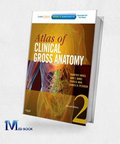
Atlas of Clinical Gross Anatomy 2nd Edition (Original PDF from Publisher)
Rated 0 out of 5
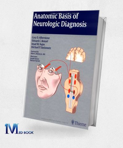
Anatomic Basis of Neurologic Diagnosis (Original PDF from Publisher)
Rated 0 out of 5
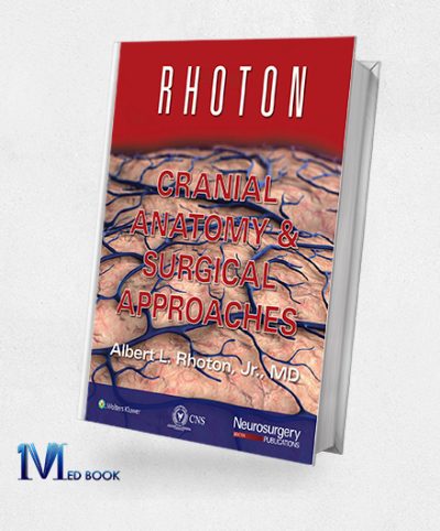
Rhoton Cranial Anatomy and Surgical Approaches (EPUB)
Rated 0 out of 5


Reviews
There are no reviews yet.