Grays Clinical Photographic Dissector of the Human Body (Original PDF from Publisher)
Grays Clinical Photographic Dissector of the Human Body (Original PDF from Publisher)
| Language |
English |
|---|---|
| Edition |
1st |
| Format |
Publisher PDF |
| ISBN-10 |
1437724175 |
| ISBN-13 |
978-1437724172, 9781437724172 |
$62.00 Original price was: $62.00.$22.00Current price is: $22.00.
- The files will be sent to you via E-mail
- Once you placed your order, we will make sure that you receive the files as soon as possible
5/5
Description
Grays Clinical Photographic Dissector of the Human Body (Original PDF from Publisher)
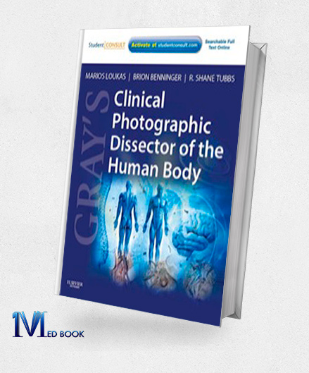
Grays Clinical Photographic Dissector of the Human Body An Authoritative Approach to Anatomical Study In the realm of anatomical exploration, the renowned publication, “Grays Clinical Photographic Dissector of the Human Body,” authored by esteemed medical professionals Drs. Marios Loukas, Brion Benninger, and R. Shane Tubbs, presents an invaluable resource tailored to a clinical perspective on anatomy.
Unlike traditional anatomical illustrations, this distinctive dissection guide employs a profusion of vibrant full-color photographs, expediting your orientation within the laboratory setting.
Every structure’s significance is underscored, coupled with an appreciation of the clinical implications inherent in each dissection conducted. Moreover, Grays Clinical Photographic Dissector of the Human Body extends its utility by elucidating contemporary emergency procedures that bear direct relevance to clinical practice, fortifying the comprehension of these critical correlations.
With an expansive assemblage of over 1,300 meticulously captured images, this compendium emerges as an exceptional conduit for acquiring, reviewing, and internalizing anatomical knowledge, intrinsically linked to its applications in the clinical realm.
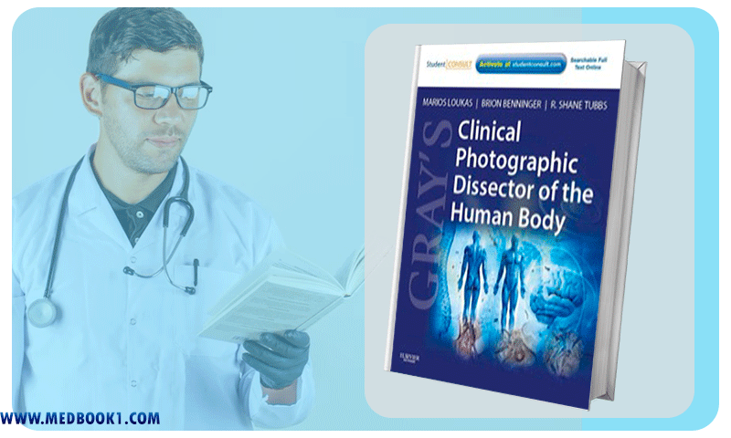
Grays Clinical Photographic Dissector of the Human Body (Original PDF from Publisher)
A hallmark of this distinguished resource is its adept facilitation of the synthesis between anatomical entities and their clinical manifestations.
This synergy empowers practitioners to navigate the intricate landscape of anatomical structures within a clinical context, thereby forging a cohesive understanding.
In the pursuit of dissection proficiency, the comprehensive compilation of 1,350 full-color photographs featured in this tome serves as an indispensable comparative tool.
By juxtaposing these vivid visual depictions with the cadavers that are subjects of study, learners can cultivate a heightened sense of assurance in their dissection endeavors.
This authoritative guide extends its purview to encompass the elucidation of 18 prevalent emergency procedures, encompassing techniques such as lumbar puncture and knee aspiration.
The meticulous exploration of these procedures equips readers with a nuanced comprehension of the anatomical substrates that underlie these interventions. Furthermore, the innate correlation between anatomical intricacies and emergent medical interventions is expounded upon, fostering a holistic grasp of clinical practice.
The caliber and meticulousness of information upheld by this resource mirrors the same rigorous standards that have etched “Gray’s Anatomy” as the quintessential reference in the realm of intricate anatomical understanding.
Dr. Marios Loukas, a luminary in the domain of clinical anatomy, infuses his expertise into the content, perpetuating the legacy of accuracy and thoroughness synonymous with the “Gray’s Anatomy” lineage.
In summation, “Grays Clinical Photographic Dissector of the Human Body” emerges as an indispensable compendium, seamlessly weaving the tapestry of anatomical comprehension with its clinical applications.
By seamlessly amalgamating the depth of anatomical structures with emergent clinical scenarios, this resource transcends traditional approaches, rendering the journey of anatomical discovery all the more enlightening and consequential.

Grays Clinical Photographic Dissector of the Human Body (Original PDF from Publisher)
1.2.Key Features
The foremost attributes that distinguish “Grays Clinical Photographic Dissector of the Human Body,” authored by Drs. Marios Loukas and Brion Benninger, encompass the following key features:
- Clinical Integration: This resource seamlessly integrates anatomical understanding with clinical relevance. Through an extensive array of full-color photographs, it fosters a profound grasp of anatomical structures within the context of real-world clinical scenarios.
- Visual Learning: Unlike traditional anatomical drawings, this guide employs over 1,350 meticulously captured photographs, allowing learners to visually compare and comprehend the intricacies of anatomical structures in relation to cadaveric specimens.
- Emergency Procedures: Gray’s Clinical Photographic Dissector of the Human Body significantly enhances learning by elucidating 18 crucial emergency procedures, including lumbar puncture and knee aspiration. It underscores the connection between anatomical knowledge and these interventions, enabling a comprehensive grasp of their clinical execution.
- Clinical Significance: Every anatomical structure is meticulously linked to its clinical importance, enabling learners to appreciate how each component translates to practical medical applications, thereby fostering a deeper understanding of medical practice.
- Authoritative Expertise: The collaboration of Drs. Marios Loukas and Brion Benninger ensures that the content reflects the highest level of clinical anatomical expertise. Dr. Loukas, an esteemed authority in the field, contributes his profound knowledge, further solidifying the guide’s credibility.
- Dissection Confidence: By providing a vast collection of full-color images for comparison to actual cadaveric specimens, the resource instills confidence in learners undertaking dissections, thereby facilitating hands-on anatomical exploration.
- Comprehensive Reference: Just as “Gray’s Anatomy” is an emblematic reference for the subject, this guide maintains the same standards of accuracy and meticulousness, offering learners a reliable and comprehensive reference for both anatomical understanding and its clinical implications.
1.3. About Writer
Drs. Marios Loukas, Brion Benninger, and R. Shane Tubbs are esteemed medical professionals who have significantly impacted the field of clinical anatomy.
Dr. Marios Loukas is a leading authority in clinical anatomy, renowned for his expertise and contributions to anatomical research and education.
His work has not only enriched anatomical understanding but also shaped clinical applications through his involvement in medical literature.
Dr. Brion Benninger has made substantial contributions to anatomical education and research, particularly in the realm of dissection techniques and clinical correlations.
His dedication to enhancing anatomical learning experiences is reflected in his contributions to educational resources like “Grays Clinical Photographic Dissector of the Human Body.”

Grays Clinical Photographic Dissector of the Human Body (Original PDF from Publisher)
Dr. R. Shane Tubbs is recognized for his pivotal role in advancing neuroanatomical knowledge.
His extensive research in neuroanatomy and neurosurgery has led to significant advancements in the understanding of the human nervous system.
His collaborative efforts have contributed to publications that bridge the gap between anatomical knowledge and clinical practice.
Collectively, these professionals have reshaped the landscape of clinical anatomy, elevating education, research, and clinical applications through their groundbreaking contributions and dedication to advancing the field.
Summary
“Grays Clinical Photographic Dissector of the Human Body,” authored by Drs. Marios Loukas, Brion Benninger, and R. Shane Tubbs, revolutionizes anatomical study through its unique approach of employing over 1,350 full-color photographs to replace traditional illustrations.
This distinctive dissection guide seamlessly bridges anatomical knowledge with clinical significance, fostering a deeper understanding of structures in practical medical contexts.
Grays Clinical Photographic Dissector of the Human Body not only aids in hands-on dissections by allowing learners to compare photographs with cadaveric specimens, but also highlights the clinical implications of each structure and introduces essential emergency procedures.
Driven by the expertise of the authors, including Dr. Loukas’ prominence in clinical anatomy, the guide sets a new standard in anatomical education, empowering learners to connect theory with real-world medical practice.
Make sure that you are buying e-books from trustworthy sources. With over a decade of experience in the e-book industry, the Medbook1.com website is a reliable option for your purchase.
Categories:
Other Products:
Genomic and Personalized Medicine 2nd Edition V1-2 (Original PDF from Publisher)
Fundamentals of Enzyme Kinetics 4th (Original PDF from Publisher)
Histology and Cell Biology An Introduction to Pathology 3rd (Original PDF from Publisher)
How to Read a Paper The Basics of Evidence Based Medicine 4th (Original PDF from Publisher)
Reviews (0)
Be the first to review “Grays Clinical Photographic Dissector of the Human Body (Original PDF from Publisher)” Cancel reply
Related products
A.D.A.M. Interactive Anatomy Online Student Lab Activity Guide 4th Edition (Original PDF from Publisher)
Rated 0 out of 5
Fields Anatomy Palpation & Surface Markings 5th Edition (Original PDF from Publisher)
Rated 0 out of 5
Clinical Neuroanatomy and Neuroscience 6th Edition (Original PDF from Publisher)
Rated 0 out of 5
Rhoton Cranial Anatomy and Surgical Approaches (EPUB)
Rated 0 out of 5
Essentials of Anatomy & Physiology 3rd edition (Original PDF from Publisher)
Rated 0 out of 5


Reviews
There are no reviews yet.