
-61%
")
Intended to infuse the process of learning with captivating engagement and clinical significance, the innovative resource titled “Netters Concise Radiologic Anatomy” seamlessly juxtaposes radiologic imagery, including advanced reconstructions, with the exquisite artwork of the esteemed medical illustrator, Dr. Frank H. Netter.
In stark contrast to other available anatomic and radiologic atlases, Netters Concise Radiologic Anatomy commences its journey from the realm of anatomy, harmonizing radiologic depictions with the anatomical renderings that have been indelibly etched into the minds of those who routinely consult Dr. Netter’s Atlas of Human Anatomy.
The accompanying succinct textual content expedites access to pivotal anatomic and radiologic insights, seamlessly entwining the visual narratives.
With a structure and chromatic arrangement mirroring the well-known Netter’s Atlas of Human Anatomy, this resource stands as an ideal companion to the atlas, offering an insightful guide to comprehending the clinical underpinnings of normal anatomy.
Within its pages, Netters Concise Radiologic Anatomy presents a side-by-side exposition of radiological instances showcasing both the normative anatomy and common variations, accompanied by corresponding anatomical illustrations.
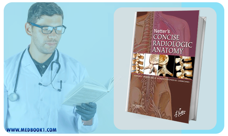
Netters Concise Radiologic Anatomy (Original PDF from Publisher)
This presentation not only underscores the clinical significance of the anatomical structures but also facilitates direct, efficient comparisons between the idealized anatomic renderings and their real-world medical counterparts.
Furthermore, Netters Concise Radiologic Anatomy incorporates a concise primer on fundamental radiological principles, including reconstructions, accompanied by a compendium of common abbreviations.
This introductory foundation fortifies the readers’ knowledge as they engage with the ensuing images.
In an effort to provide a seamless continuity of reference, Netters Concise Radiologic Anatomy adheres to the familiar organizational structure and color-coded scheme utilized in Netter’s Atlas of Human Anatomy.
This congruity streamlines cross-referencing and reinforces the familiarity that users have cultivated with Dr. Netter’s iconic style.
Editorial feedback attests to the resource’s value within medical education. One review highlights its utility as an adjunct to gross anatomy studies, particularly beneficial to students with an affinity for radiology or surgical disciplines.
While the resource does not aim to supplant traditional anatomic atlases or comprehensive radiologic references, its innovative utilization of image reconstruction facilitates a smooth transition from illustrative anatomical portrayals to the complexities of modern radiological image interpretation.
The authors’ intention of acquainting anatomy scholars with radiologic images and illuminating how contemporary radiology captures human anatomy is undoubtedly realized.
However, it is worth noting that a foundational understanding of human gross anatomy is presupposed for effective utilization of this atlas.
As such, the atlas stands as a valuable addition to the repertoire of anatomical reference materials, finding particular resonance among medical students during clerkships and residents who seek to rekindle their anatomical insights while correlating them with radiological representations.
The commendable quality of Netter’s artwork is aptly juxtaposed with the excellence of the corresponding radiological images, thus effectively fulfilling the atlas’s mission of harmonizing superior radiological visuals with Netter’s anatomical depictions.
This modern, high-caliber radiological atlas is poised to captivate an audience comprising medical students, allied health professionals, surgical and radiology residents, and the broader cohort of hospital-based practitioners.
Amidst these deliberations, an interesting observation surfaces from a distinct anecdote. In December 2009, a report emerges from an unrelated field, tangentially invoking “Netters Concise Radiologic Anatomy.”
Within this context, a patient’s medical history intertwines with the atlas’s narrative, creating an unforeseen connection that underscores the resource’s enduring presence in the medical domain.
Netters Concise Radiologic Anatomy
The most prominent key features of “Netters Concise Radiologic Anatomy” are as follows:
Dr. Frank H. Netter, MD (1906–1991), was a prolific American medical illustrator and a trailblazer in the field of medical artistry.
Renowned for his exceptional ability to translate complex medical concepts into visually comprehensible illustrations, he left an indelible mark on medical education.
Graduating from New York University School of Medicine in 1931, Netter’s career flourished as he combined his medical knowledge with his artistic prowess.
His magnum opus, “Atlas of Human Anatomy,” published in 1989, is celebrated as a cornerstone of medical education, praised for its clarity, accuracy, and clinical relevance.
Netter’s intricate depictions transformed medical learning, making anatomy accessible to generations of students, physicians, and allied health professionals.
His work extended beyond anatomy, encompassing pathology, physiology, and clinical medicine.
Dr. Netter’s artistic legacy continues to illuminate medical education, reflecting his lifelong commitment to bridging the gap between art and science.
His legacy endures through the ongoing impact of his artwork on medical curricula worldwide, forever enriching the understanding of the human body.
Netters Concise Radiologic Anatomy (Original PDF from Publisher)
“Netters Concise Radiologic Anatomy” represents a groundbreaking educational resource that seamlessly combines the intricate radiologic images, including advanced reconstructions, with the iconic anatomical illustrations of Dr. Frank H. Netter.
This unique atlas facilitates a direct comparison between clinical radiological depictions and the well-established anatomical renderings, providing a comprehensive understanding of normal anatomy and its clinical relevance.
Following the familiar structure of Netter’s Atlas of Human Anatomy, Netters Concise Radiologic Anatomy side-by-side presentations and concise text empower learners, from medical students to practicing doctors, with the ability to swiftly access key anatomical and radiological insights.
With its fusion of artistry and medical imaging, this resource serves as an invaluable companion guide, bridging the gap between traditional anatomical understanding and modern radiological interpretation.
Make sure that you are buying e-books from trustworthy sources. With over a decade of experience in the e-book industry, the Medbook1.com website is a reliable option for your purchase.
Categories:
Other Products:
Netters Atlas of Human Embryology Updated Edition 1st (Netter Basic Science)
Nanomaterials in Drug Delivery Imaging and Tissue Engineering (Original PDF from Publisher)
Muscle Biopsy A Practical Approach 4th Edition (Original PDF from Publisher)
Netters Head and Neck Anatomy for Dentistry 2nd Edition (Netter Basic Science)
No account yet?
Create an Account


Reviews
There are no reviews yet.