Histology
Atlas of Radiologic Cytopathologic Correlations (Original PDF from Publisher)
Original price was: $115.00.$22.00Current price is: $22.00.Biopsy Interpretation of the Central Nervous System (Biopsy Interpretation Series)
Original price was: $154.85.$26.00Current price is: $26.00.Biopsy Interpretation The Frozen Section 2nd Edition
Original price was: $139.93.$28.00Current price is: $28.00.Cellular Imaging Techniques for Neuroscience and Beyond (Original PDF from Publisher)
Original price was: $118.92.$26.60Current price is: $26.60.Concise Histology With STUDENT CONSULT Online Access (Original PDF from Publisher)
Original price was: $45.40.$22.40Current price is: $22.40.Cytology Diagnostic Principles and Clinical Correlates 4th Edition (ORIGINAL PDF from Publisher)
Original price was: $176.64.$29.70Current price is: $29.70.Diagnostic Cytopathology Essentials (Original PDF from Publisher)
Original price was: $156.36.$28.60Current price is: $28.60.Diagnostic Cytopathology Expert Consult Online and Print 3rd Edition (Original PDF from Publisher)
Original price was: $291.65.$36.00Current price is: $36.00.Elseviers Integrated Histology (Original PDF from Publisher)
Original price was: $63.26.$23.40Current price is: $23.40.Essentials of Oral Histology and Embryology A Clinical Approach 4th Edition (Original PDF from Publisher)
Original price was: $72.62.$24.00Current price is: $24.00.Extreme Tissue Engineering Concepts and Strategies for Tissue Fabrication (Original PDF from Publisher)
Original price was: $57.00.$21.00Current price is: $21.00.Histology A Text and Atlas With Correlated Cell and Molecular Biology, 9th Edition (EPUB)
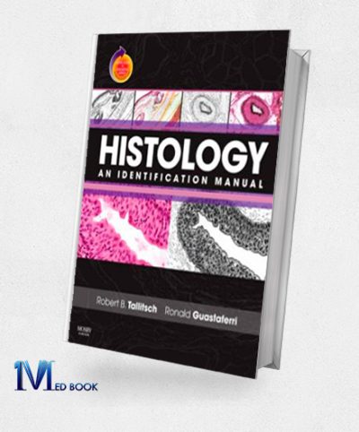
Original price was: $113.99.$48.00Current price is: $48.00.Histology An Identification Manual (Original PDF from Publisher)
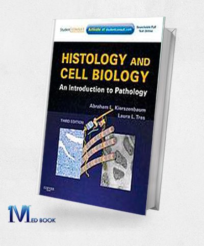
Original price was: $54.76.$22.00Current price is: $22.00.Histology and Cell Biology An Introduction to Pathology 3rd (Original PDF from Publisher)
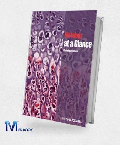
Original price was: $66.00.$21.00Current price is: $21.00.Histology at a Glance (Original PDF from Publisher)
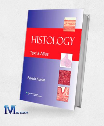
Original price was: $38.51.$21.90Current price is: $21.90.Histology Text and Atlas (Brijesh Kumar)
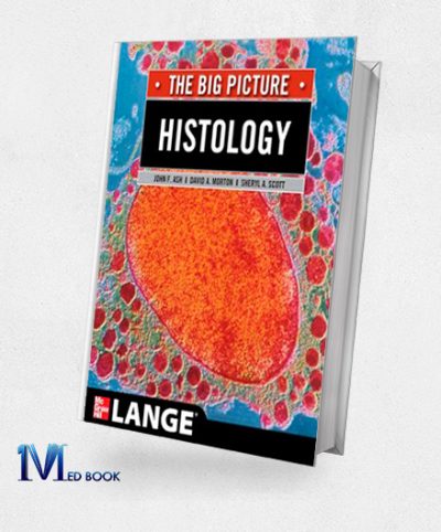
Original price was: $33.21.$22.00Current price is: $22.00.Histology The Big Picture (LANGE The Big Picture) (EPUB)
Original price was: $42.75.$21.60Current price is: $21.60.Intraoperative Frozen Sections Diagnostic Pitfalls (Consultant Pathology) (Original PDF from Publisher)
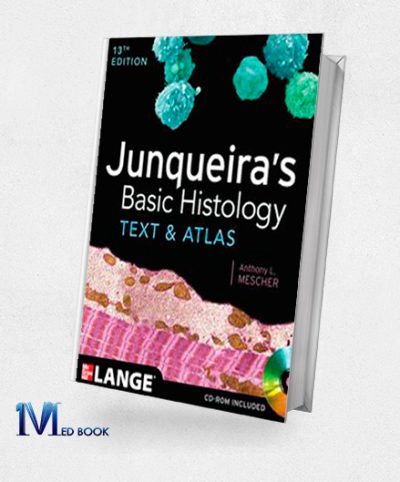
Original price was: $164.99.$21.00Current price is: $21.00.Junqueiras Basic Histology Text and Atlas 13th Edition (Original PDF from Publisher)
Original price was: $58.88.$23.00Current price is: $23.00.Lange Basic Histology Flash Cards (Original PDF from Publisher)
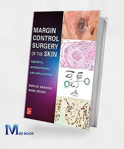
Original price was: $34.82.$23.00Current price is: $23.00.Margin Control Surgery of the Skin Concepts Histopathology and Applications (EPUB)
Original price was: $89.00.$30.00Current price is: $30.00.Margin Control Surgery of the Skin Concepts Histopathology and Applications (Original PDF from Publisher)
Original price was: $89.00.$30.00Current price is: $30.00.Histology
Introduction to Histology
Histology, the microscopic study of tissues and their structures serves as a fundamental discipline in unraveling the intricate organization and function of tissues within living organisms. At its core, histology delves into the microscopic realms, allowing us to explore the cellular architecture that forms the building blocks of life. By examining tissues at the microscopic level, histology provides invaluable insights into the composition, arrangement, and dynamic interactions of cells within various organs and structures.
The significance of histology extends beyond mere observation, as it plays a crucial role in deciphering the physiological processes, identifying abnormalities, and understanding the harmonious collaboration of different tissues that sustain life. Whether investigating the delicate intricacies of organs or elucidating the structural nuances of diverse tissues, histology serves as a gateway to comprehending the marvels of cellular biology. Through this lens, we embark on a journey to unravel the mysteries of living organisms at a scale invisible to the naked eye, shedding light on the profound intricacies that govern the functioning and unity of life.
Types of Tissues Studied in Histology
In the realm of histology, the study encompasses a diverse array of tissues, each with distinctive characteristics and specialized functions. Epithelial tissue, forming the protective covering of organs and surfaces, serves as a barrier against external elements. Its stratified or simple structure reflects adaptations tailored to specific locations, such as the skin or inner linings.
Connective tissue is a versatile and supportive category that binds and sustains various structures within the body. It plays a pivotal role in maintaining structural integrity, ranging from the dense framework of bones to the pliability of tendons.
Muscle tissue powers movement and locomotion with its contractile abilities. Skeletal muscles, which are under voluntary control, orchestrate bodily motions, while smooth and cardiac muscles contribute to involuntary functions, such as digestive processes and heart contractions.
Nervous tissue is an intricate network of neurons and supporting cells that forms the foundation of the nervous system. By facilitating communication through electrical impulses, nervous tissue enables sensory perception, motor responses, and intricate cognitive functions.The exploration of these tissue types in histology provides a profound understanding of the harmonious interplay that underlies the intricate machinery of the human body. By unraveling the unique characteristics and functions of epithelial, connective, muscle, and nervous tissues, histology contributes to the comprehensive comprehension of the complex tapestry of life at the microscopic level.
Microscopic Techniques in Histology
Histology, the study of tissues at the microscopic level, relies on a myriad of sophisticated techniques to unravel the intricacies of cellular structures. Staining methods serve as a cornerstone, imparting distinct colors to tissues and cells, and enhancing their visibility under microscopes. This aids in distinguishing various components, such as nuclei and cytoplasm, facilitating a comprehensive analysis.
Light microscopy, a fundamental tool in histology, employs visible light to illuminate specimens. This technique enables the observation of overall tissue architecture and cellular arrangements. It is particularly valuable for examining stained sections and obtaining a broad understanding of tissue organization.
Electron microscopy is an advanced approach that uses electron beams to investigate smaller details. Transmission electron microscopy (TEM) can provide ultra-high resolution, revealing intricate subcellular structures such as organelles and membranes. Scanning electron microscopy (SEM) offers a three-dimensional view, which can help to better understand surface features.
Immunohistochemistry is another vital technique that involves labeling specific proteins with antibodies. This technique can help researchers to pinpoint the localization of proteins within tissues, which in turn contributes to a deeper understanding of cellular functions and interactions.
Confocal microscopy is known for its optical sectioning ability, which enables the reconstruction of three-dimensional images. This technique is particularly valuable for studying complex tissues and visualizing cellular relationships in depth.
In summary, the arsenal of microscopic techniques in histology, including staining methods, light microscopy, electron microscopy, immunohistochemistry, and confocal microscopy, collectively empowers researchers to explore tissues at unparalleled resolutions. These techniques play a pivotal role in advancing our understanding of cellular structures and their intricate relationships within the tapestry of living organisms.
Cellular Components and Structures in Histology
Histology delves into the microscopic study of cellular components and structures, unraveling the intricacies of tissues and their organizational patterns. At the core of histological exploration is the understanding of cellular organization within tissues. Tissues, composed of groups of specialized cells, collaborate to perform specific functions essential for the overall health of an organism.
Within individual cells, various organelles contribute to cellular functions. The nucleus, housing genetic material, regulates cellular activities and serves as the command center. The endoplasmic reticulum, Golgi apparatus, and vesicles collectively participate in protein synthesis, modification, and transportation. Mitochondria, the powerhouse of the cell, generate energy through cellular respiration.
Examining tissues in histology reveals distinctive cellular arrangements. Epithelial tissues, forming protective layers or lining organs, showcase tightly packed cells. Connective tissues, such as bone or blood, display diverse cell types embedded in an extracellular matrix. Muscle tissues exhibit elongated cells that contract for movement, while nervous tissues contain neurons for transmitting electrical impulses.
Histological analysis discerns the significance of specific structures in the context of tissue function. For instance, cilia and microvilli on epithelial cells contribute to the absorption and movement of substances. In muscle tissues, the organization of sarcomeres facilitates contraction. The intricate capillary network within tissues ensures efficient nutrient exchange.
In essence, histology illuminates the nuanced architecture of tissues, highlighting the organization of cells and the pivotal role of cellular structures in facilitating tissue-specific functions. This microscopic exploration is indispensable for comprehending the building blocks of living organisms and the orchestration of cellular activities that sustain life.
Functional Correlations in Histology
Histology intricately weaves the tapestry of living organisms by exploring the profound relationship between the microscopic features of tissues and their physiological functions. At the heart of this discipline is the recognition that the structure of tissues profoundly influences their function, forming the basis for the harmonious orchestration of life processes.
Epithelial tissues are composed of tightly packed cells that have specialized structures such as cilia and microvilli. These tissues line organs and surfaces, and they serve different functions such as absorption, secretion, or acting as barriers. These functions are intricately linked to the tissue’s cellular arrangement.
Connective tissues, on the other hand, are made up of diverse cell types embedded in an extracellular matrix. This matrix composition, whether in bone, blood, or cartilage, determines the tissue’s flexibility, resilience, or transport capabilities. Connective tissues play a crucial role in supporting and connecting various body components.
Muscle tissues are composed of organized sarcomeres and contractile proteins that facilitate precise and coordinated movements. These movements are vital for bodily functions such as locomotion and organ function.
Nervous tissues are composed of neurons and supporting cells that transmit electrical impulses. The complex architecture of neurons, including axons and dendrites, enables rapid communication, forming the basis of nervous system function.
Histology’s exploration of functional correlations extends beyond individual tissues to unravel the intricate networks within organs. Understanding how the microscopic features contribute to the physiological functions of organs is essential in grasping the complexity of living organisms.
In essence, histology serves as a gateway to deciphering the remarkable interplay between structure and function in living organisms. By unveiling the microscopic intricacies of tissues, it provides profound insights into the mechanisms that sustain life and the fascinating harmony between form and function.
Histopathology and Disease Diagnosis
Histopathology is a crucial tool in diagnosing diseases. It involves examining tissues at a microscopic level to identify abnormalities, which helps pathologists identify the nature and characteristics of diseases. This information is instrumental in determining appropriate treatment strategies and predicting prognosis.
Histopathology is also useful in assessing disease progression. By analyzing tissues, pathologists can understand the stage and severity of the condition, which helps in devising effective treatment plans.
Moreover, histopathology plays a significant role in guiding treatment decisions. The microscopic examination of tissues provides vital information to multidisciplinary teams, helping them select the most appropriate therapeutic interventions for patients.
Overall, histopathology is an essential aspect of disease diagnosis and treatment, offering critical insights into the microscopic realm that can help improve patient outcomes.informs on the potential responsiveness of the disease to specific treatments, enabling a personalized and targeted approach.
The integration of histopathology into disease diagnosis is a testament to its profound impact on clinical decision-making. Its ability to unveil the hidden signatures of diseases at the cellular level empowers healthcare professionals with the knowledge needed for accurate diagnosis, prognosis, and treatment planning. In essence, histopathology serves as a microscopic detective, deciphering the intricate narratives written within tissues to illuminate the path toward effective disease management.
Histology in Research and Medical Education
Histology plays a multifaceted role in both research and medical education, serving as a cornerstone for scientific advancements, medical training, and deepening our understanding of disease mechanisms. In research, histological studies provide a microscopic lens into the intricate structures and functions of tissues, facilitating breakthroughs in various scientific disciplines. By enabling researchers to visualize cellular details, histology contributes crucial insights into normal physiology and pathological processes. This knowledge forms the basis for innovative discoveries, advancements in medical treatments, and a deeper comprehension of biological phenomena.
In medical education, histology serves as an indispensable tool for training the next generation of healthcare professionals. Medical students learn about the microscopic anatomy of tissues, understand the intricacies of cellular organization, and how these structures relate to physiological functions. Histological slides become educational resources, allowing students to bridge the gap between textbook knowledge and the practical application of their understanding. Through hands-on experiences with histological specimens, medical students develop essential diagnostic skills and gain insights into the manifestations of diseases at the cellular level.
Histology is particularly instrumental in unraveling disease mechanisms. By examining tissues microscopically, researchers and medical professionals can identify abnormalities, study the progression of diseases, and uncover key pathological features. This knowledge is pivotal in developing targeted therapeutic strategies and refining diagnostic approaches. Histology thus stands at the forefront of medical advancements, guiding research endeavors that contribute to improved patient care and outcomes.
In essence, histology’s applications in research and medical education are intertwined, creating a symbiotic relationship that fuels scientific progress and enhances the quality of healthcare. Its role in dissecting the microscopic intricacies of tissues ensures that the medical community continues to build on a foundation of knowledge that is essential for advancing both the understanding of disease and the training of future healthcare practitioners.
Emerging Technologies in Histology
Histology, the study of tissues at the microscopic level, is undergoing a transformative phase with the integration of emerging technologies that enhance capabilities and provide novel insights. One such innovation is digital pathology, which involves the digitization of histological slides. This technology allows pathologists and researchers to view and analyze specimens remotely, fostering collaboration and easing the exchange of information. Digital pathology not only streamlines workflows but also facilitates the integration of artificial intelligence for automated analysis, potentially expediting diagnoses.
Another groundbreaking development is three-dimensional imaging in histology. Traditional histological techniques produce two-dimensional representations, but three-dimensional imaging offers a more comprehensive view of tissue structures. This advancement provides a better understanding of the spatial relationships between cells and enhances the visualization of complex anatomical features. It has particular relevance in fields such as developmental biology and neuroscience, where the three-dimensional organization of tissues is critical.
Molecular histology is a cutting-edge approach that combines traditional histological methods with molecular biology techniques. This allows researchers to investigate the molecular composition of tissues alongside their microscopic structures. By analyzing specific biomolecules within cells, molecular histology provides insights into the molecular processes underlying normal physiology and disease pathology. This integration of molecular and histological information contributes to a more holistic understanding of tissue function and dysfunction.
These emerging technologies collectively push the boundaries of what is achievable in histology, offering researchers and pathologists unprecedented tools for exploration. The integration of digital pathology, three-dimensional imaging, and molecular histology not only refines diagnostic approaches but also opens new avenues for uncovering the intricate details of tissue biology. As these technologies continue to evolve, they hold the promise of revolutionizing histological research and further advancing our understanding of the microscopic world within living organisms.
Challenges and Future Directions in Histology
Histology, despite its pivotal role in understanding tissues at the microscopic level, faces several challenges that warrant consideration for future development. One of the primary challenges is the standardization of techniques across laboratories and institutions. Variability in histological protocols and practices can lead to inconsistencies in results, hindering the reproducibility of studies. Addressing this challenge involves establishing standardized protocols and promoting adherence to best practices within the histological community.
Ethical considerations in histology represent another important aspect. As technologies advance, issues related to the use of human or animal tissues for research purposes come to the forefront. Balancing the need for scientific progress with ethical concerns about tissue sourcing, consent, and privacy requires ongoing discussions and the formulation of guidelines to ensure responsible and transparent practices.
The integration of histological data with findings from other disciplines is a key area for future exploration. Bridging the gap between histology and fields such as genomics, proteomics, and bioinformatics could provide a more comprehensive understanding of tissue biology. Efforts to develop interdisciplinary collaborations and establish common frameworks for data integration will be crucial for maximizing the utility of histological information in the broader context of biomedical research.
In addition, the advent of digital pathology and big data in histology presents both opportunities and challenges. Managing and analyzing large datasets generated through high-throughput techniques requires advanced computational tools and infrastructure. Striking a balance between harnessing the power of big data in histology and ensuring data security, quality, and interpretation poses a complex challenge.
Looking ahead, the future of histology holds promise in overcoming these challenges through collaborative efforts, technological innovations, and ethical considerations. By addressing issues related to standardization, ethics, and interdisciplinary integration, the field can continue to evolve, providing valuable insights into the intricate world of tissues and contributing significantly to biomedical research and clinical applications.
Conclusion
Histology is a fundamental discipline that helps us understand the complexities of tissue structures, and it plays a crucial role in advancing our knowledge of health and disease. By examining tissues at the microscopic level, histological studies provide unparalleled insights into their cellular and architectural composition, which is essential for comprehending how the human body functions normally and identifying any deviations associated with various diseases.
Histology is a cornerstone in medical research and education, and it contributes to the development of diagnostic and therapeutic strategies. Healthcare professionals can use histology to make accurate diagnoses, assess disease progression, and guide effective treatment decisions. Beyond its clinical applications, histology fosters continuous advancements in medical knowledge, laying the foundation for innovations in fields ranging from pathology to regenerative medicine.
As we navigate the complexities of health and disease, histology remains an invaluable tool that illuminates the intricate tapestry of cellular structures. Its significance extends beyond the confines of laboratories, impacting the broader landscape of medical science. By emphasizing the essential role of histology, we acknowledge its enduring contribution to the enhancement of our understanding of the human body, ultimately promoting advancements in healthcare for the benefit of individuals and society as a whole.

