
-61%
")
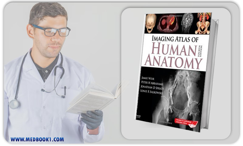
The fourth edition of the “Imaging Atlas of Human Anatomy” serves as a comprehensive foundational resource for comprehending the intricate details of human anatomical structures.
Authored by Jamie Weir, Peter Abrahams, Jonathan D. Spratt, and Lonie Salkowski, this edition presents a comprehensive three-dimensional depiction of bodily structures and their interrelationships, utilizing a diverse range of imaging techniques.
With the inclusion of over 60% new images, including cross-sectional views from CT and MRI scans, nuclear medicine imaging, and other modalities, coupled with revised captions and annotations, this atlas offers an exceptionally current and top-tier visual reference.
Moreover, readers gain access to a set of 10 pathology tutorials online, with the option to purchase an additional 24 tutorials, which provide links to supplementary images, thereby offering an even more exhaustive coverage than before.
Both in its physical form and online counterpart, this atlas significantly enriches applied and clinical understanding of human anatomy.
The distinctive attributes of this edition include orientation drawings that aid in comprehending diverse perspectives and orientations within the images, accompanied by tables detailing the timeline of bone development.
The images are meticulously labeled with numerical identifiers, preserving their clarity and facilitating self-assessment. Legends and labels have undergone a complete revision, and an impressive majority of the new images, surpassing 60%, encompass cross-sectional views obtained from CT and MRI scans, as well as angiography, ultrasound, fetal anatomy, plain film anatomy, and nuclear medicine imaging.
These images feature heightened resolution, rendering them the most current and precise anatomical representations available.
The revised content accurately mirrors contemporary radiological and anatomical practices, exemplified by the restructured chapters on the abdominal and pelvic regions, which now include a novel chapter dedicated to cross-sectional imaging.
Encompassing a wide spectrum of prevalent and modern imaging techniques, including a freshly introduced section on Nuclear Medicine, this edition offers insights into living anatomical structures, thereby enhancing comprehension based on dissection and artistic representations.
The incorporation of static representations of three-dimensional images further aids in grasping dynamic visual concepts. Additionally, readers can access ten pathology tutorials free of charge online. These tutorials, collaboratively developed with recent medical students, are accompanied by numerous pathological images, augmenting the development of visual memory pertaining to anatomical structures and their spatial orientations.
In essence, the “Imaging Atlas of Human Anatomy 4th” stands as an authoritative repository of knowledge,
providing a profound understanding of human anatomy through a myriad of cutting-edge imaging techniques. The frequent incorporation of the keyword “Imaging Atlas of Human Anatomy 4th” within the context aligns with the thematic essence of the atlas, underscoring its significance and relevance.
Imaging Atlas of Human Anatomy 4th (Original PDF from Publisher)
The paramount features of this book include:
Imaging Atlas of Human Anatomy
Jamie Weir, a prominent figure in the field of anatomical education, is an esteemed author of the “Imaging Atlas of Human Anatomy 4th Edition.”
With a profound understanding of medical imaging and anatomy, Weir has made significant contributions to anatomical education, enhancing the comprehension of complex structures through visual representation.
Peter H. Abrahams, an accomplished anatomist and co-author, has played a pivotal role in shaping medical education.
His expertise in anatomical dissection and imaging has been instrumental in creating a comprehensive and clinically relevant atlas.
Abraham’s commitment to advancing anatomical knowledge is evident through his numerous publications and teaching engagements.
Jonathan D. Spratt, an esteemed author and anatomist, has enriched anatomical education through his contributions to the atlas.
His expertise in radiological anatomy and cross-sectional imaging has added a contemporary dimension to the atlas, reflecting his commitment to aligning anatomical education with modern medical practices.
Lonie R. Salkowski, a valued collaborator, brings a wealth of expertise in medical illustration and anatomical visualization to the team.
Salkowski’s contributions have enhanced the visual clarity and educational impact of the atlas, making intricate anatomical structures more accessible to learners.
Together, these authors have shaped the “Imaging Atlas of Human Anatomy 4th Edition,” fostering a deeper understanding of human anatomy through their collective achievements in anatomical education, imaging, and medical illustration.
Imaging Atlas of Human Anatomy 4th (Original PDF from Publisher)
The “Imaging Atlas of Human Anatomy 4th Edition,” authored by Jamie Weir, Peter H. Abrahams, Jonathan D. Spratt, and Lonie R Salkowski, stands as a comprehensive and contemporary resource offering a multidimensional view of human anatomical structures through diverse imaging modalities.
With over 60% new images, including cross-sectional views from CT and MRI scans, nuclear medicine imaging, and more, the atlas provides a clear and up-to-date visual reference.
Noteworthy features encompass orientation drawings, numerical labeling for self-testing, and tables of bone development timelines.
The inclusion of online pathology tutorials, coupled with a focus on clinical relevance, further enriches readers’ understanding of anatomical structures and their practical implications.
This collaborative effort reflects the authors’ expertise in anatomical education, radiological anatomy, and medical illustration, making the atlas an invaluable tool for both learners and healthcare professionals seeking to deepen their anatomical knowledge.
Make sure that you are buying e-books from trustworthy sources.With over a decade of experience in the e-book industry, the Medbook1.com website is a reliable option for your purchase.
Categories:
Other Products:
Introduction to Healthcare Information Technology

Koda Kimble and Youngs Applied Therapeutics The Clinical Use of Drugs 10th Edition

Lab Experiences for the Pharmacy Technician 2nd Edition
No account yet?
Create an Account

Reviews
There are no reviews yet.