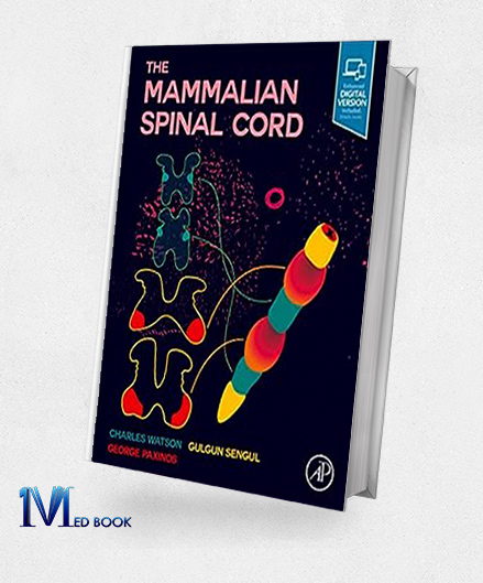
The Mammalian Spinal Cord (Original PDF from Publisher)
The Mammalian Spinal Cord (Original PDF from Publisher)
$180.00 Original price was: $180.00.$45.00Current price is: $45.00.
- The files will be sent to you via E-mail
- Once you placed your order, we will make sure that you receive the files as soon as possible
The Mammalian Spinal Cord (Original PDF from Publisher)
1.1.Description
“The Mammalian Spinal Cord” is an exhaustive study focusing on the intricate anatomy and histology of this crucial neurological structure. Encompassing cytoarchitecture, chemoarchitecture, motor neuron distribution, long tracts, autonomic outflow, and gene expression, it stands as an indispensable resource for spinal cord researchers.
Notably, it features segment-by-segment atlases showcasing the spinal cords of rats, mice, newborn mice, marmosets, rhesus monkeys, and humans, incorporating vivid full-color photographic Nissl-stained sections. Accompanying these are meticulously labeled diagrams, enhancing the understanding of each segment.
Moreover, the book presents over 500 photographic images of sections stained for various markers, augmenting the comprehensive Nissl-stained images with details on AChE, ChAT, parvalbumin, NADPH-diaphorase, calretinin, and others.”

The Mammalian Spinal Cord (Original PDF from Publisher)
1.2.Key Features
The key features of “The Mammalian Spinal Cord” include:
- Comprehensive Anatomy: Provides an extensive account of spinal cord anatomy and histology, covering cytoarchitecture, chemoarchitecture, motor neuron distribution, long tracts, autonomic outflow, and gene expression.
- Segment-by-Segment Atlases: “The Mammalian Spinal Cord” Includes detailed atlases displaying spinal cords from various species (rat, mouse, newborn mouse, marmoset, rhesus monkey, and human) with full-color photographic Nissl-stained sections, aiding comprehensive understanding.
- Detailed Diagrams: Accompanies each Nissl-stained section with over 160 comprehensively labeled diagrams, facilitating a clearer interpretation of the intricate spinal cord structures.
- Supplementary Images: “The Mammalian Spinal Cord” Offers more than 500 photographic images of sections stained for different markers (AChE, ChAT, parvalbumin, NADPH-diaphorase, calretinin, etc.), enhancing the understanding beyond Nissl-stained images.
- Research Reference: This serves as an essential reference for researchers and scholars exploring the complexities of the spinal cord, making it a fundamental resource in the field of neuroscience.

The Mammalian Spinal Cord (Original PDF from Publisher)
1.3. About Writer
Charles Watson, Gulgun Sengul, and George Paxinos are distinguished figures in the field of neuroscience. George Paxinos, a renowned neuroscientist, is notably recognized for his significant contributions to brain cartography, having created the Paxinos-Watson atlas series, an extensive collection of brain atlases widely used by neuroscientists globally.
Paxinos collaborated with Charles Watson, an esteemed researcher and neuroanatomist, on numerous publications related to brain mapping and neuroanatomy, contributing significantly to the field’s advancement. Gulgun Sengul, another prominent figure in neuroscience, has been an integral part of Paxinos’s team, actively involved in the production and enhancement of brain atlases and neuroanatomical research.
Together, their collective work has profoundly influenced the understanding of brain structure and function, providing invaluable resources for neuroscientists, clinicians, and researchers worldwide.

The Mammalian Spinal Cord (Original PDF from Publisher)
Summary
“The Mammalian Spinal Cord” serves as an extensive resource detailing the intricate anatomy and histology of the spinal cord. This comprehensive text covers various aspects, including cytoarchitecture, chemoarchitecture, motor neuron distribution, long tracts, autonomic outflow, and gene expression within the spinal cord.
Noteworthy features of the book encompass segment-by-segment atlases of the spinal cords from different species, such as rodents like rats, mice, newborn mice, marmosets, rhesus monkeys, and humans. The inclusion of full-color photographic images of Nissl-stained sections, labeled diagrams, and supplementary images stained for various markers enhances the understanding of spinal cord anatomy and makes this book an indispensable reference for researchers delving into spinal cord studies.


Reviews
There are no reviews yet.