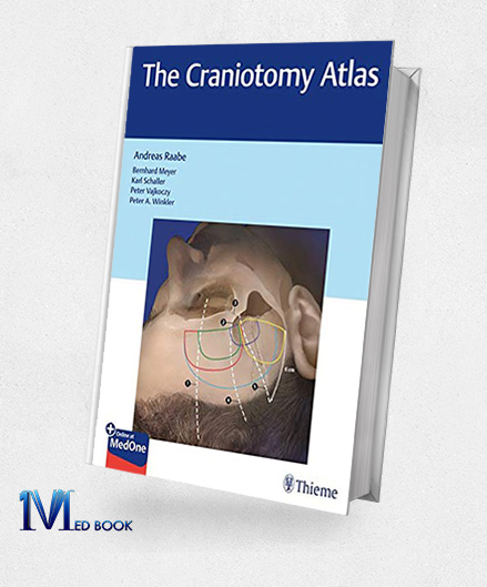
The Craniotomy Atlas (PDF)
The Craniotomy Atlas (PDF)
| Edition |
1 |
|---|---|
| File Size |
44.4 MB |
| Format |
Publisher PDF |
| Language |
English |
| Publisher |
Thieme |
| ISBN-10 |
9783132057913 |
$189.11 Original price was: $189.11.$25.00Current price is: $25.00.
- The files will be sent to you via E-mail
- Once you placed your order, we will make sure that you receive the files as soon as possible
5/5
Description
The Craniotomy Atlas (PDF)
1.1.Description
Mastering the craniotomy procedure is crucial for junior residents, given that the majority of brain surgeries commence with this step. The ability to select the appropriate craniotomy and execute it safely distinguishes a straightforward case with optimal access to the target from a potentially traumatic and challenging procedure.
In “The Craniotomy Atlas (PDF)” Professor Raabe offers precise instructions for a range of common neurosurgical cranial exposures, encompassing convexity approaches, midline approaches, skull base approaches, transsphenoidal approaches, and more.
The comprehensive guide includes detailed information on positioning, head fixation, aesthetic considerations, and dura mater protection for each craniotomy.
With over 600 high-quality operative photographs and illustrations supporting step-by-step descriptions, this resource, edited by Professor Raabe and associate editors Professors Meyer, Schaller, Vajkoczy, and Winkler, ensures the precision and attention to detail expected by neurosurgeons.
The book also covers complications, and risk factors, and features a checklist summarizing critical steps. This essential reference, accompanied by complimentary digital access, instills confidence in residents and trainees undertaking critical neurosurgical procedures.

The Craniotomy Atlas
1.2.Key Features
“The Craniotomy Atlas (PDF)” stands out for its key features that make it an essential resource for junior residents and trainees in neurosurgery:
- Comprehensive Coverage: “The Craniotomy Atlas (PDF)” provides precise instructions for performing common neurosurgical cranial exposures, including convexity approaches, midline approaches, skull base approaches, and transsphenoidal approaches.
- Operative Visuals: More than 600 high-quality operative photographs and brilliant illustrations support step-by-step descriptions, offering visual clarity and guidance for each craniotomy procedure.
- Editorial Expertise: Edited by Professor Raabe, along with associate editors Professors Meyer, Schaller, Vajkoczy, and Winkler, the atlas ensures precision and attention to detail, reflecting the expertise of renowned neurosurgeons.
- Risk Factors and Complications: Full coverage of complications and risk factors associated with craniotomies provides valuable insights for practitioners, enhancing their understanding and preparedness.
- Checklist for Critical Steps: The atlas includes a checklist summarizing critical steps, offering a quick reference for practitioners to ensure procedural accuracy and safety.
- Aesthetic Considerations: Instructions for each craniotomy include aesthetic considerations, addressing the importance of appearance in addition to procedural precision.
- Digital Access: “The Craniotomy Atlas (PDF)” includes complimentary access to a digital copy on https://medone.thieme.com, enhancing accessibility and convenience for residents and trainees.
- Confidence Building: Aimed at building confidence among junior residents and trainees, the atlas serves as an essential guide for performing critical neurosurgical procedures.
In summary, “The Craniotomy Atlas (PDF)” combines detailed procedural guidance, visual support, editorial expertise, and digital accessibility to provide a comprehensive and confidence-building resource for neurosurgical practitioners.

The Craniotomy Atlas
Summary
“The Craniotomy Atlas (PDF)” is an indispensable resource designed to empower junior residents and trainees in neurosurgery with precise instructions for common neurosurgical cranial exposures.
Edited by Professor Raabe and renowned associate editors, including Professors Meyer, Schaller, Vajkoczy, and Winkler, the atlas encompasses a comprehensive range of craniotomy procedures, from convexity and midline approaches to skull base and transsphenoidal approaches.
With over 600 high-quality operative photographs and illustrations providing visual clarity, the atlas ensures a step-by-step guide to each procedure, addressing crucial aspects such as positioning, head fixation, aesthetic considerations, and dura mater protection.
Covering complications, and risk factors, and featuring a checklist for critical steps, the atlas combines precision with accessibility.
Complimentary digital access further enhances its utility, making it an essential confidence-building reference for residents and trainees undertaking critical neurosurgical procedures.
Reviews (0)
Be the first to review “The Craniotomy Atlas (PDF)” Cancel reply
Related products
Schmidek and Sweet Operative Neurosurgical Techniques 2-Volume Set Indications, Methods and Results, 7th Edition (Original PDF from Publisher)
Rated 0 out of 5
Cranial Neuroimaging And Clinical Neuroanatomy Atlas Of MR Imaging And Computed Tomography, 4th Edition (ORIGINAL PDF From Publisher)
Rated 0 out of 5
Anatomy Vivas for the Intercollegiate MRCS (Original PDF from Publisher)
Rated 0 out of 5
Anatomy for Anaesthetists 9th Edition (Original PDF from Publisher)
Rated 0 out of 5
Uropathology A Volume in the High Yield Pathology Series (Original PDF from Publisher)
Rated 0 out of 5



Reviews
There are no reviews yet.