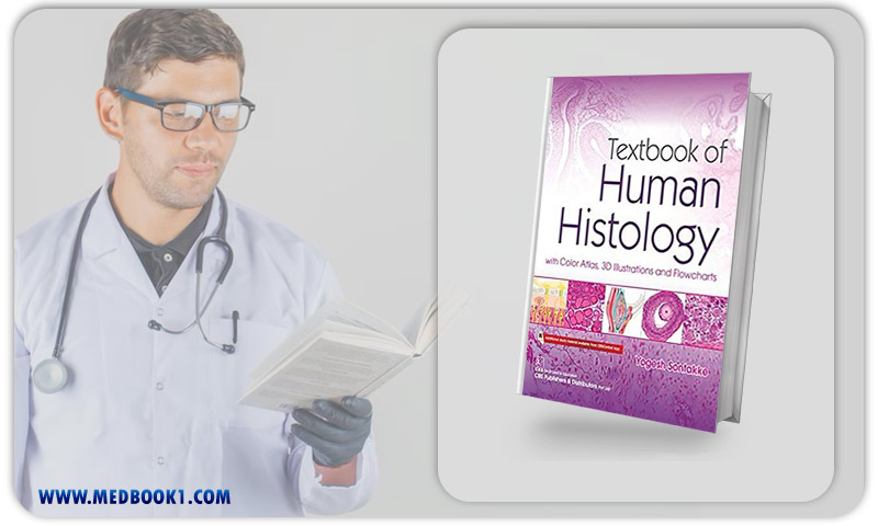
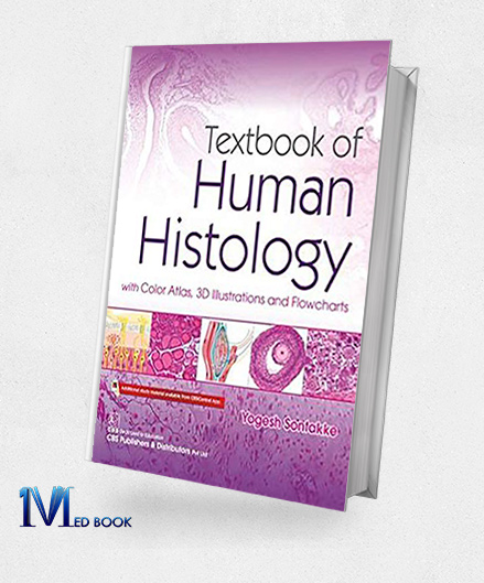
Textbook of Human Histology With Color Atlas 3D Illustrations and Flowcharts (Original PDF from Publisher)
Textbook of Human Histology With Color Atlas 3D Illustrations and Flowcharts (Original PDF from Publisher)
| Edition |
1st |
|---|---|
| Format |
Publisher PDF |
| ISBN-10 |
9788194125426 |
| Language |
English |
| Publisher |
Other Publisher |
$70.17 Original price was: $70.17.$24.00Current price is: $24.00.
- The files will be sent to you via E-mail
- Once you placed your order, we will make sure that you receive the files as soon as possible
5/5
Description
Textbook of Human Histology With Color Atlas 3D Illustrations and Flowcharts (Original PDF from Publisher)
1.1.Description
“Textbook of Human Histology With Color Atlas 3D Illustrations and Flowcharts” on human histology is designed for undergraduate medical students, offering concise text with functional correlations for efficient review during examinations. The content is enriched with 117 photomicrographs, aiding in the identification of microscopic structures, along with 122 flowcharts that facilitate quick revision and memorization of microanatomy concepts.
To support theoretical examinations, there are 106 practice figures in the form of HE pencil drawings that can be easily reproduced. The inclusion of 175 3D illustrations provides students with a visual understanding of challenging concepts. A summary, serving as an examination guide, addresses the challenge of summarizing facts in written assessments.
Additionally, the textbook incorporates interesting facts, set apart from the main text to ensure they are not overlooked, and offers clinical correlations to orient students toward the pathogenesis of diseases, promoting vertical integration.

Textbook of Human Histology With Color Atlas 3D Illustrations and Flowcharts (Original PDF from Publisher)
1.2.Key Features
The key features of “Textbook of Human Histology With Color Atlas 3D Illustrations and Flowcharts” include a concise text with functional correlations for efficient exam preparation, 117 photomicrographs for identifying microscopic structures, 122 flowcharts for quick revision, and 106 practice figures for easy reproduction in theory exams.
The textbook also incorporates 175 3D illustrations for a visual grasp of complex concepts, a summary for effective examination preparation, interesting facts to enhance engagement, and clinical correlations that provide insight into the pathogenesis of diseases. This comprehensive resource aims to support undergraduate medical students in mastering human histology.

Textbook of Human Histology With Color Atlas 3D Illustrations and Flowcharts (Original PDF from Publisher)
Summary
“Textbook of Human Histology With Color Atlas 3D Illustrations and Flowcharts” is a comprehensive resource designed for undergraduate medical students, offering a thorough understanding of human histology. The textbook presents concise text with functional correlations, aiding quick recapitulation during examinations.
With 117 photomicrographs, 122 flowcharts, and 106 practice figures, the book facilitates visual learning and revision of microanatomy. The inclusion of 175 3D illustrations provides a unique and detailed visual grasp of complex concepts. Additionally, a summary (examination guide) is provided to assist students in summarizing essential facts for written assessments.
“Textbook of Human Histology With Color Atlas 3D Illustrations and Flowcharts” incorporates interesting facts, clinical correlations, and vertical integration to enhance the overall learning experience in histology.
Reviews (0)
Be the first to review “Textbook of Human Histology With Color Atlas 3D Illustrations and Flowcharts (Original PDF from Publisher)” Cancel reply


Reviews
There are no reviews yet.