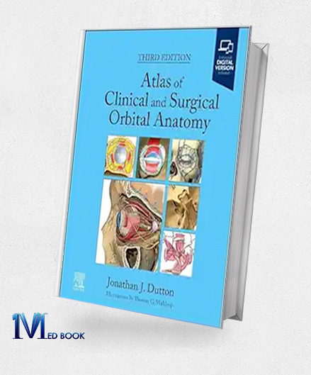
Atlas Of Clinical And Surgical Orbital Anatomy, 3rd Edition (True PDF)
Atlas Of Clinical And Surgical Orbital Anatomy, 3rd Edition (True PDF)
| Edition |
3rd |
|---|---|
| File Size |
170.5 MB |
| Format |
True PDF (No Index) |
| ISBN-10 |
0443109427 |
| ISBN-13 |
978-0443109423, 9780443109652, 9780443109669 |
| Language |
English |
| Publisher |
Elsevier |
$251.99 Original price was: $251.99.$60.00Current price is: $60.00.
- The files will be sent to you via E-mail
- Once you placed your order, we will make sure that you receive the files as soon as possible
5/5
Description
Atlas Of Clinical And Surgical Orbital Anatomy, 3rd Edition (True PDF)
1.1.Description
For surgeons and ophthalmologists, the orbit—a complicated bone cavity that houses the eye and its related structures—presents a special difficulty. A useful tool for understanding this complex anatomy is Dr. Jonathan J. Dutton’s Atlas of Clinical and Surgical Orbital Anatomy, Third Edition (True PDF).
This thorough atlas is more than just a representation of features; it is a fundamental resource for comprehending the intricate details of the clinical and surgical orbital anatomy.
Traditional anatomy texts are surpassed by this painstakingly created atlas of clinical and surgical ocular anatomy.
Dr. Dutton’s work is a valuable resource for ophthalmologists, oculoplastic surgeons, neurosurgeons, and any other medical professional working with the orbit since it provides a distinctive synthesis of excellent graphics, thorough text descriptions, and perceptive clinical linkages.

Atlas Of Clinical And Surgical Orbital Anatomy, 3rd Edition (True PDF)
An grasp of orbital embryology is fundamental to the atlas of clinical and surgical orbital anatomy. The foundation for understanding the intricate developmental processes that form the orbit is laid forth in this section.
The adult orbital anatomy is covered in detail in later chapters, along with descriptions of the bones, muscles, nerves, blood vessels, and other important components that are contained inside the orbit.
Dr. Dutton’s skillful use of pictures is what makes him so strong. An abundance of finely detailed, multi-layered anatomical artwork may be found in the Atlas of Clinical and Surgical Orbital Anatomy. These drawings provide unmatched clarity and depth by showing the orbit in a variety of planes and dissections.
Understanding the complex interactions between different orbital structures is essential for understanding clinical and surgical orbital anatomy, and this graphic method makes it easier for readers to do so.
The text continues with thorough descriptions that flow naturally from the illustrations. Dr. Dutton ensures that the function and significance of each anatomical feature are fully understood by giving succinct and understandable explanations of each. This combination of text and images makes the complex orbital architecture easier to understand.

Atlas Of Clinical And Surgical Orbital Anatomy, 3rd Edition (True PDF)
However, the emphasis on clinical correlations in this atlas of clinical and surgical orbital anatomy is what really makes it valuable. Dr. Dutton skillfully closes the gap between clinical anatomy knowledge and its practical application.
Every chapter carefully examines the therapeutic ramifications of particular anatomical characteristics. For example, the text talks about how different orbital bone shapes might affect surgical techniques or how distinct ophthalmic illnesses can be explained by particular nerve pathways.
The atlas of clinical and surgical orbital anatomy is elevated above a simple anatomical reference by its emphasis on clinical relationships. It gives readers the information and understanding they need to manage the complexity of orbital surgery and make wise clinical judgments.
The third edition has a number of notable improvements. Comprehensive discussion of the anatomy and clinical relevance of the cavernous sinus, a crucial orbital structure, is offered in a newly devoted chapter.
To guarantee that readers have access to the most recent knowledge, the drawings have also been painstakingly updated to reflect the most recent anatomical discoveries.
Additionally, a related website is accessible through the atlas of clinical and surgical ocular anatomy.
This online resource offers a treasure trove of downloadable images, allowing readers to seamlessly incorporate the illustrations into presentations or teaching materials.

Atlas Of Clinical And Surgical Orbital Anatomy, 3rd Edition (True PDF)
To sum up, Dr. Jonathan J. Dutton’s Atlas of Clinical and Surgical Orbital Anatomy, Third Edition (True PDF) is a tribute to the careful research of orbital anatomy.
This atlas, with its superb graphics, thorough text explanations, and illuminating clinical connections, is an invaluable tool for neurosurgeons, ophthalmologists, oculoplastic surgeons, and any other medical professional who wants to learn more about the orbit.
By enabling readers to successfully navigate the complexity of orbital structures, this thorough guide will eventually improve surgical outcomes and patient care.
Make sure that you are buying e-books from trustworthy sources. With over a decade of experience in the e-book industry, the Medbook1.com website is a reliable option for your purchase.
Categories:
Other Products:
| Professional Communication in Audiology (Original PDF from Publisher) |
| The Temporal Bone Anatomical Dissection And Surgical Approaches (PDF) |
Audiology Science To Practice, 4th Edition (Original PDF From Publisher)
Audiology Workbook, 4th Edition (Original PDF From Publisher)
Reviews (0)
Be the first to review “Atlas Of Clinical And Surgical Orbital Anatomy, 3rd Edition (True PDF)” Cancel reply
Related products
General Organic and Biochemistry 11th edition (Original PDF from Publisher)
Rated 0 out of 5
Biology Laboratory Manual 13th edition (Original PDF from Publisher)
Rated 0 out of 5
Anesthesia Oral Board Review 2nd edition (Original PDF from Publisher)
Rated 0 out of 5
2024 2025 Saunders Clinical Judgment and Test Taking Strategies 8th edition (Original PDF from Publisher)
Rated 0 out of 5
Neuroanatomy through Clinical Cases 3rd edition (Original PDF from Publisher)
Rated 0 out of 5






Reviews
There are no reviews yet.