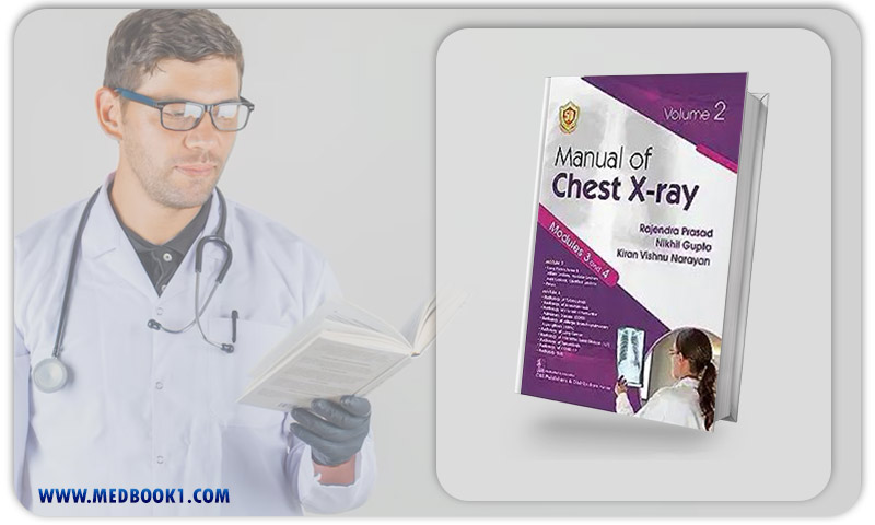
Manual of Chest X-ray, Volume 2 ( Modules 3 and 4 ) (Original PDF from Publisher)
Manual of Chest X-ray, Volume 2 ( Modules 3 and 4 ) (Original PDF from Publisher)
$44.00 Original price was: $44.00.$30.00Current price is: $30.00.
- The files will be sent to you via E-mail
- Once you placed your order, we will make sure that you receive the files as soon as possible
Manual of Chest Xray Volume 2 ( Modules 3 and 4 ) (Original PDF from Publisher)
1.1.Description
This comprehensive manual on chest X-ray interpretation, titled “Manual of Chest Xray Volume 2 (Modules 3 and 4)” offers a thorough examination of various aspects of chest X-ray analysis.
It encompasses a systematic approach to reading normal chest X-rays and identifying lesions in different anatomical areas, including soft tissue, bony cage, diaphragm, hilum, heart, mediastinum, lung parenchyma, and pleura.
The content is organized systematically for easy comprehension and is available in both print and e-book formats, divided into four modules across two volumes.
Module 3 covers lesions such as miliary, nodular, mass, calcified, and various pleural lesions, while Module 4 delves into chest X-ray findings related to tuberculosis, bronchiectasis, COPD, sarcoidosis, interstitial lung diseases, lung cancer, allergic bronchopulmonary Aspergillosis, and Covid-19.
This manual is valuable for medical students, residents, radiologists, physicians, and surgeons engaged in the day-to-day interpretation of chest X-rays in clinical practice.

Manual of Chest Xray Volume 2
1.2.Key Features
The key features of “Manual of Chest Xray Volume 2 (Modules 3 and 4)” include:
- Comprehensive Coverage: The manual provides a thorough examination of chest X-ray interpretation, covering normal chest X-rays and various lesions in different anatomical structures.
- Systematic Approach: The content is organized systematically, facilitating easy understanding and systematic reading of chest X-rays.
- Module Structure: “Manual of Chest Xray Volume 2 (Modules 3 and 4)” is divided into four modules across two volumes. Modules 3 and 4 specifically cover lesions such as miliary, nodular, mass, calcified, and pleural lesions, and various chest X-ray findings related to specific medical conditions.
- Clinical Relevance: The content is clinically relevant, making it useful for medical students, residents, radiologists, physicians, and surgeons involved in the interpretation of chest X-rays in day-to-day clinical practice.
- Available Formats: “Manual of Chest Xray Volume 2 (Modules 3 and 4)” is available in both print and e-book versions, providing flexibility for different learning preferences.
- Specialized Content: Module 4 includes discussions on chest X-ray findings related to tuberculosis, bronchiectasis, COPD, sarcoidosis, interstitial lung diseases, lung cancer, allergic bronchopulmonary Aspergillosis, and COVID-19.
These features make “Manual of Chest Xray Volume 2 (Modules 3 and 4)” a valuable resource for individuals at various stages of their medical careers involved in interpreting chest X-rays.

Manual of Chest Xray Volume 2
1.3. About Writer
Dr. Rajendra Prasad, Dr. Nikhil Gupta, and Dr. Kiran Vishnu Narayan are esteemed medical professionals with significant achievements in their respective fields.
Dr. Rajendra Prasad is renowned for his expertise in diagnostic radiology, specializing in imaging techniques and interpretation. Dr. Nikhil Gupta has made substantial contributions to the field of pulmonology, focusing on respiratory disorders and advanced diagnostic methods.
Dr. Kiran Vishnu Narayan has excelled in the domain of cardiology, with a special emphasis on cardiovascular imaging and intervention.
Together, their collaborative efforts have likely led to advancements in the interdisciplinary approach to medical diagnostics and treatment.
Their collective contributions contribute to the progress of medical knowledge and the enhancement of patient care in their respective specialties.

Manual of Chest Xray Volume 2
Summary
“Manual of Chest Xray Volume 2 (Modules 3 and 4)” serves as a comprehensive guide to chest X-ray interpretation, covering various aspects systematically.
This manual, presented in two volumes, explores the systemic reading of normal chest X-rays and delves into lesions in soft tissue, bony cage, diaphragm, hilum, heart, mediastinum, lung parenchyma, and pleura.
The third module covers military, nodular, mass, calcified, and various pleural lesions, while the fourth module discusses chest X-ray findings related to tuberculosis, bronchiectasis, COPD, sarcoidosis, interstitial lung diseases, lung cancer, allergic bronchopulmonary aspergillosis, and COVID-19.
The organization of chapters facilitates easy understanding, making it a valuable resource for medical students, residents, radiologists, physicians, and surgeons engaged in chest X-ray interpretation in clinical practice.


Reviews
There are no reviews yet.