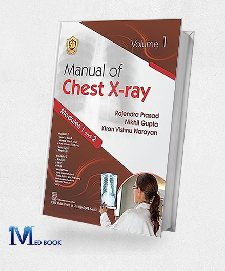
Manual of Chest X-ray, Volume 1 ( Modules 1 and 2 ) (Original PDF from Publisher)
Manual of Chest X-ray, Volume 1 ( Modules 1 and 2 ) (Original PDF from Publisher)
$44.00 Original price was: $44.00.$30.00Current price is: $30.00.
- The files will be sent to you via E-mail
- Once you placed your order, we will make sure that you receive the files as soon as possible
Manual of Chest Xray Volume 1 ( Modules 1 and 2 ) (Original PDF from Publisher)
1.1.Description
“Manual of Chest Xray Volume 1 (Modules 1 and 2)” stands as a comprehensive guide to chest X-ray interpretation, encompassing all facets of this diagnostic practice.
This manual, available in both print and e-book versions, is divided into four modules across two volumes, systematically organized for easy comprehension.
Module 1 covers the reading of normal chest X-rays and lesions in soft tissues, bony cages, and diaphragms. Module 2 explores lesions of the heart, mediastinum, hilum, cavity, and infiltrations.
Catering to medical students, residents, radiologists, physicians, and surgeons engaged in the day-to-day interpretation of chest X-rays, this manual serves as a valuable resource for understanding normal and abnormal findings in diverse chest structures and conditions.

Manual of Chest Xray Volume 1
1.2.Key Features
The key features of “Manual of Chest Xray Volume 1 (Modules 1 and 2)” include:
- Comprehensive Coverage: The manual provides a thorough examination of chest X-ray interpretation, addressing normal findings and various lesions across different chest structures.
- Systematic Organization: The content is systematically organized into modules, with Volume 1 covering Module 1 (normal chest X-rays and lesions in soft tissues, bony cage, and diaphragm) and Module 2 (lesions of heart, mediastinum, hilum, cavity, and infiltrations).
- Print and E-book Versions: “Manual of Chest Xray Volume 1 (Modules 1 and 2)” is available in both print and e-book formats, offering flexibility in accessing the content.
- Audience Inclusivity: Tailored for a diverse audience, including medical students, residents, radiologists, physicians, and surgeons involved in the daily practice of interpreting chest X-rays.
- User-Friendly Design: The systematic organization and clear presentation make the manual user-friendly, facilitating easy understanding of chest X-ray interpretations.
- Two-Volume Structure: “Manual of Chest Xray Volume 1 (Modules 1 and 2)” is divided into two volumes, each covering specific modules, providing a focused and organized approach to chest X-ray education.
- Clinical Relevance: “Manual of Chest Xray Volume 1 (Modules 1 and 2)” emphasizes clinical relevance, making it a valuable resource for professionals engaged in day-to-day clinical practice.

Manual of Chest Xray Volume 1
1.3. About Writer
Rajendra Prasad, Nikhil Gupta, and Kiran Vishnu Narayan are esteemed authors who have made significant contributions to the field of radiology and medical education.
Dr. Rajendra Prasad brings a wealth of expertise in radiology, with a focus on chest imaging. His research and clinical experience have contributed to the advancement of diagnostic practices in the interpretation of chest X-rays.
Dr. Nikhil Gupta, with a background in radiology, has demonstrated a commitment to education and clinical excellence. His collaborative efforts in producing educational resources reflect his dedication to enhancing the knowledge and skills of medical professionals.
Dr. Kiran Vishnu Narayan, as a co-author, has likely played a pivotal role in creating comprehensive manuals that bridge the gap between theoretical understanding and practical application in chest X-ray interpretation.
Together, their collective achievements underscore their commitment to advancing medical education and improving patient care in the field of radiology.

Manual of Chest Xray Volume 1
Summary
“Manual of Chest Xray Volume 1 (Modules 1 and 2)” is a comprehensive guide to chest X-ray interpretation, covering essential aspects of normal and pathological findings.
Organized into modules, the first volume addresses systemic readings of normal chest X-rays and lesions involving soft tissues, bony cage, and diaphragm (Module 1).
The second module delves into lesions related to the heart, mediastinum, hilum, cavity, and infiltrations (Module 2).
The book offers a systematic approach for easy comprehension, making it an invaluable resource for medical students, residents, radiologists, physicians, and surgeons involved in the day-to-day clinical interpretation of chest X-rays.
With a focus on clarity and accessibility, this manual serves as an essential tool for enhancing the skills of healthcare professionals in the field of radiology, providing a robust foundation for accurate and informed diagnostic practices.


Reviews
There are no reviews yet.