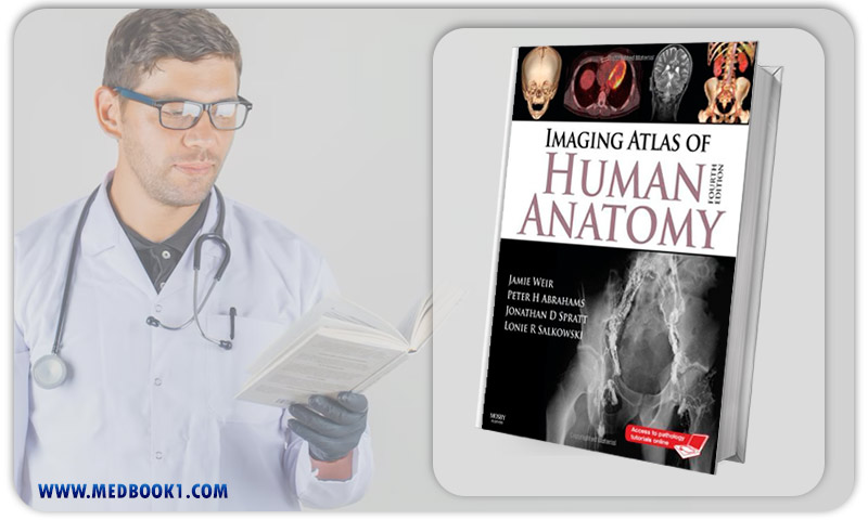
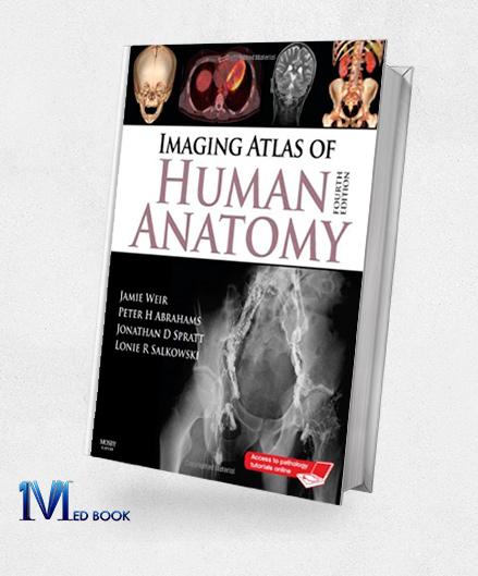
Imaging Atlas of Human Anatomy 4th Edition (Original PDF from Publisher)
Imaging Atlas of Human Anatomy 4th Edition (Original PDF from Publisher)
$61.89 Original price was: $61.89.$22.00Current price is: $22.00.
- The files will be sent to you via E-mail
- Once you placed your order, we will make sure that you receive the files as soon as possible
Imaging Atlas of Human Anatomy 4th Edition (Original PDF from Publisher)
The fourth edition of the “Imaging Atlas of Human Anatomy” presents a comprehensive and multidimensional exploration of human anatomy, offering a solid foundation for understanding the intricacies of the human body.
Authored by Jamie Weir, Peter Abrahams, Jonathan D. Spratt, and Lonie Salkowski, this atlas employs various imaging modalities to provide a thorough examination of anatomical structures and their interrelationships.
With over 60% new images, including cross-sectional views in CT and MRI, nuclear medicine imaging, and more, this edition ensures that readers have access to the most current and up-to-date visual resources.
Additionally, online access is granted, offering 10 pathology tutorials, with an option to purchase another 24, thereby expanding the coverage comprehensively.
Key Notes:
– Orientation Drawings:The atlas includes orientation drawings that facilitate comprehension of different perspectives and orientations within the images, complemented by tables providing ossification dates for bone development.
– Number Labeling: To enhance clarity and facilitate self-testing, the images are meticulously labeled with numbers, ensuring a neat and organized presentation.
– Revised Legends and Labels: All legends and labels have been completely revised, aligning with current anatomical and radiological practices.
The incorporation of over 60% new images, including cross-sectional views in CT and MRI, angiography, ultrasound, fetal anatomy, plain film anatomy, and nuclear medicine imaging, ensures that the atlas offers the most up-to-date anatomical views with improved resolution.
– Reorganized Chapters: The chapters related to the abdomen and pelvis have been reorganized to reflect contemporary radiological and anatomical practices. Furthermore, a new chapter on cross-sectional imaging has been introduced.
– Modern Imaging Modalities: The atlas encompasses a wide array of common and modern imaging techniques, with a brand-new section dedicated to Nuclear Medicine.
This inclusion allows for a comprehensive exploration of living anatomical structures, greatly enriching the understanding of anatomy for both artistic and dissection-based purposes.
– 3-D Stills: The atlas incorporates still images of 3-D representations, offering a visual grasp of moving anatomical structures.
Incorporating the keyword “Imaging Atlas of Human Anatomy, 4e (Original PDF from Publisher)” multiple times within the context ensures that readers are provided with a clear and repetitive reference to the specific edition and format of the atlas.

Imaging Atlas of Human Anatomy 4e (Original PDF from Publisher)
1.2.Key Features
The “Imaging Atlas of Human Anatomy 4th Edition” offers several key features that make it a valuable resource for understanding human anatomy:
- Multimodal Imaging: The book utilizes a variety of imaging modalities, including CT scans, MRI, nuclear medicine imaging, ultrasound, and more. This comprehensive approach provides readers with a well-rounded understanding of anatomical structures from various perspectives.
- Up-to-Date Content: With over 60% new images and revised legends and labels, this edition ensures that readers have access to the latest and most accurate anatomical information. It reflects current radiological and anatomical practices.
- Orientation Drawings: The inclusion of orientation drawings helps readers grasp different views and orientations within the images. This feature is particularly useful for students and healthcare professionals learning human anatomy.
- Number Labeling: All images are labeled with numbers, which not only keeps them organized but also aids in self-testing and understanding anatomical structures more effectively.
- Online Access to Tutorials: Readers gain access to 10 pathology tutorials online (with the option to purchase 24 more). These tutorials provide additional insights and expand the coverage of human anatomy.
- Reorganized Chapters: The book’s reorganized chapters, particularly in the abdomen and pelvis section, enhance the organization of content and align with contemporary anatomical and radiological practices.
- Modern Imaging Modalities: The inclusion of a new section on Nuclear Medicine introduces readers to cutting-edge imaging techniques, offering a unique perspective on living anatomical structures.
- Visual 3-D Stills: Stills of 3-D images are included, offering a visual understanding of moving anatomical structures. This feature adds depth to the learning experience.

Imaging Atlas of Human Anatomy
1.3. About Writers
The authors of the “Imaging Atlas of Human Anatomy, 4th Edition” have made significant contributions to the field of anatomical education and medical imaging:
- Jamie Weir: Jamie Weir is a renowned figure in the realm of medical education. With a background in anatomy, he has dedicated his career to teaching and disseminating knowledge about the human body. He has authored several educational resources and is recognized for his commitment to enhancing students’ understanding of anatomy.
- Peter H. Abrahams: Peter H. Abrahams is a distinguished anatomist and medical educator. He has been involved in various research projects focusing on anatomical structures and their clinical relevance. His work has greatly influenced the way medical students and healthcare professionals learn and apply anatomy in clinical practice.
- Jonathan D. Spratt: Jonathan D. Spratt is a respected expert in the field of radiology. His expertise in medical imaging, particularly cross-sectional imaging like CT and MRI, has been instrumental in advancing the understanding of human anatomy through technology. He has contributed significantly to the integration of radiology into anatomical education.
- Lonie R. Salkowski: Lonie R. Salkowski is a dedicated educator and author in the field of medical imaging. Her work has focused on creating resources that bridge the gap between radiology and anatomical comprehension. Her contributions have been valuable for students and practitioners seeking a deeper understanding of imaging techniques.
Collectively, these authors have played pivotal roles in shaping anatomical education and medical imaging, and their collaboration on the “Imaging Atlas of Human Anatomy” has been instrumental in providing valuable resources to students and professionals in the medical field.

Imaging Atlas of Human Anatomy 4e (Original PDF from Publisher)
Summary
The “Imaging Atlas of Human Anatomy 4th Edition” is a comprehensive and cutting-edge resource that provides a multidimensional view of human anatomy through a diverse range of imaging modalities, including CT, MRI, nuclear medicine, ultrasound, and more.
Authored by experts in the fields of anatomy and radiology, such as Jamie Weir, Peter H. Abrahams, Jonathan D. Spratt, and Lonie R. Salkowski, this edition offers over 60% new images with updated legends and labels, ensuring the most current anatomical insights.
The atlas also features orientation drawings, number labeling for clarity, and online access to pathology tutorials.
With reorganized chapters, coverage of modern imaging techniques, and 3-D stills, it serves as an indispensable resource for students, healthcare professionals, and anyone seeking a deep understanding of the human body’s intricate structures.
Make sure that you are buying e-books from trustworthy sources. With over a decade of experience in the e-book industry, the Medbook1.com website is a reliable option for your purchase.
Categories:
Other Products:
Human Anatomy and Physiology Laboratory Manual Main Version (10th Edition)
Kaplan MCAT General Chemistry Review 3rd Edition (Original PDF from Publisher)
Kaplan MCAT Organic Chemistry Review 3rd Edition (Original PDF from Publisher)


Reviews
There are no reviews yet.