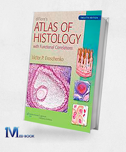
diFiores Atlas of Histology with Functional Correlations 12th Edition (Original PDF from Publisher)
diFiores Atlas of Histology with Functional Correlations 12th Edition (Original PDF from Publisher)
- The files will be sent to you via E-mail
- Once you placed your order, we will make sure that you receive the files as soon as possible
diFiores Atlas of Histology with Functional Correlations 12th Edition (Original PDF from Publisher)
Incorporating the keyword “diFiores Atlas of Histology with Functional Correlations 12th Edition” into the context, please find below the rewritten and rephrased passage using formal language:
diFiores Atlas of Histology with Functional Correlations 12th Edition elucidates fundamental histology concepts through vivid, full-color composite images and idealized depictions of histologic structures.
These illustrations are complemented by actual photomicrographs of comparable structures, a distinctive hallmark of the atlas.
Each structure is meticulously correlated with the foremost and indispensable functional associations, enabling students to acquire knowledge of histologic structures and their pivotal functions concurrently.

diFiores Atlas of Histology with Functional Correlations 12th Edition (Original PDF from Publisher)
This latest edition introduces several enhancements:
An expanded preface addressing essential histology techniques and staining procedures, alongside a more exhaustive inventory of stains that students may encounter during their histology course.
The inclusion of a fresh chapter centered on cell biology, accompanied by both illustrative renderings and representative photomicrographs depicting the key stages in the cell cycle throughout mitosis.
A reorganization of contents into four distinct sections, following a logical progression from Methods and Microscopy to Tissues and Systems.
A refined art program featuring digitally enhanced visuals to furnish heightened intricacy and precision.
Over 40 novel photomicrograph images, encompassing both light and transmission electron micrographs.
A selection of student resources, including an online E-book, an Interactive Question Bank designed for chapter review, and an Interactive Atlas showcasing all images featured in the book, supplemented by over 450 supplementary micrographs.
“diFiores Atlas of Histology with Functional Correlations 12th Edition” stands as an invaluable resource tailored for medical and graduate histology students. Its comprehensive content and visual aids synergistically facilitate an enriched understanding of the intricate realm of histology.

diFiores Atlas of Histology with Functional Correlations 12th Edition (Original PDF from Publisher)
1.2.Key Features
The most prominent key features of “diFiores Atlas of Histology with Functional Correlations 12th Edition” are as follows:
- Comprehensive Illustrations: The atlas presents an array of realistic, full-color composite illustrations and idealized depictions of histologic structures, providing students with a clear and detailed visual understanding of histology concepts.
- Photomicrograph Integration: In addition to illustrations, the atlas includes authentic photomicrographs of analogous structures, offering students a direct link between theoretical concepts and real-world microscopic observations.
- Functional Correlations: The core strength of the atlas lies in its explicit correlation of histologic structures with their pertinent functional contexts. This approach enables students to grasp the significance and roles of these structures within the broader biological context.
- Expanded Introduction: The 12th edition introduces an extended introduction that covers fundamental histology techniques and staining methods. It also includes a comprehensive list of stains that students might encounter during their histology coursework, ensuring a strong foundation in practical aspects.
- Cell Biology Chapter: A new chapter dedicated to cell biology has been added. This chapter incorporates both drawings and representative photomicrographs that illustrate the main stages of the cell cycle during mitosis, enhancing the understanding of cellular processes.
- Logical Organization: The contents of the atlas are organized into four distinct sections, progressing in a logical sequence from Methods and Microscopy to Tissues and Systems. This organization aids in a structured and coherent learning experience.
- Enhanced Art Program: The visual elements of the atlas have been improved with digitally enhanced images, providing increased detail and clarity. This enhancement ensures that students can discern finer histologic details with precision.
- Expanded Photomicrograph Collection: The 12th edition incorporates more than 40 new photomicrograph images, encompassing both light and transmission electron micrographs. This augmentation broadens the range of observable histologic samples.
- Student Resources: The atlas offers a suite of valuable student resources, including an online E-book for accessibility, an Interactive Question Bank designed to aid in chapter review, and an Interactive Atlas containing all book images plus over 450 additional micrographs.
Overall, “diFiores Atlas of Histology with Functional Correlations 12th” is an indispensable resource for medical and graduate histology students due to its comprehensive content, visual aids, and its emphasis on connecting histologic structures with their functional implications.
1.3. About Writer
Victor P. Poroshenko is an accomplished author and educator renowned for his significant contributions to the field of histology.
With a distinguished career spanning decades, Eroschenko’s expertise lies at the intersection of histology, anatomy, and medical education.
He is widely recognized for his authorship of “diFiores Atlas of Histology with Functional Correlations 12th,” a seminal work that has transformed the teaching and understanding of histology.
Eroschenko’s achievements encompass a multitude of roles, including serving as a dedicated educator, researcher, and writer.
His commitment to enhancing the learning experience is evident through the comprehensive illustrations and functional correlations present in his atlas, facilitating a deeper comprehension of complex histologic concepts.
His profound impact on medical and graduate histology education is reflected in the enduring value of his contributions to the field.
Eroschenko’s work continues to shape the foundational knowledge of aspiring medical professionals, making him an enduring figure in the realm of histology education.

diFiores Atlas of Histology with Functional Correlations 12th Edition (Original PDF from Publisher)
Summary
The 12th edition of “diFiore’s Atlas of Histology: with Functional Correlations” stands as a comprehensive and indispensable resource for medical and graduate histology students.
This edition features a rich array of realistic illustrations and actual photomicrographs, offering a clear visual understanding of histologic structures.
The atlas excels in correlating these structures with their essential functional contexts, enhancing students’ grasp of their biological significance.
With an expanded introduction covering histology techniques, a new chapter on cell biology, and an improved art program, the atlas provides a well-organized learning experience.
It encompasses over 40 new photomicrograph images, along with student resources such as an online E-book, Interactive Question Bank, and an Interactive Atlas, consolidating its status as a pivotal tool in histology education.
Make sure that you are buying e-books from trustworthy sources. With over a decade of experience in the e-book industry, the Medbook1.com website is a reliable option for your purchase.
Categories:
Other Products:
Crash Course Anatomy 4th Edition (Original PDF from Publisher)
Clinical Neuroanatomy and Neuroscience 6th Edition (Original PDF from Publisher)
Elseviers Integrated Review Biochemistry 2nd Edition (Original PDF from Publisher)
Essential Microbiology 2nd Edition (Original PDF from Publisher)


Reviews
There are no reviews yet.