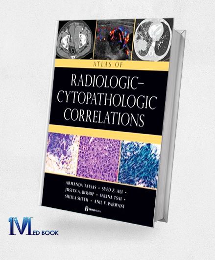
Atlas of Radiologic Cytopathologic Correlations (Original PDF from Publisher)
Atlas of Radiologic Cytopathologic Correlations (Original PDF from Publisher)
$115.00 Original price was: $115.00.$22.00Current price is: $22.00.
- The files will be sent to you via E-mail
- Once you placed your order, we will make sure that you receive the files as soon as possible
Atlas of Radiologic Cytopathologic Correlations (Original PDF from Publisher)
The “Atlas of Radiologic Cytopathologic Correlations” emerges as an indispensable resource, crucial for achieving precise and accurate interpretations of pathologic processes through the amalgamation of radiology and cytopathology.
This meticulously crafted atlas encompasses a generous compilation of over 700 meticulously chosen, high-resolution images from both radiology and cytopathology, catering to the diagnostic intricacies associated with deep-seated mass lesions, while also extending coverage to selected regions within soft tissues, bones, and superficial sites such as the thyroid.
Organized across seven chapters, this atlas facilitates effortless correlation and juxtaposition of radiologic and pathologic images, furnishing a comprehensive visual guide that encompasses all pivotal facets of radiology, cytopathology, and histopathology across key disease processes inherent to each organ system.
Within its pages, readers will encounter an array of exceptional features, including 749 high-resolution images from radiology, cytopathology, and histopathology meticulously arranged for intuitive correlation, an expansive scope that encompasses various organ systems and disease processes, an inclusive approach that spans both non-neoplastic and benign lesions in addition to malignancy, and the esteemed authorship of expert faculty representing both diagnostic specialties, culminating in a harmonious fusion of radiology and cytopathology expertise.

Atlas of Radiologic Cytopathologic Correlations (Original PDF from Publisher)
This “Atlas of Radiologic Cytopathologic Correlations” stands as a beacon of knowledge, providing invaluable guidance to practitioners and learners navigating the complexities of pathological diagnosis through a comprehensive integration of visual cues and clinical insights.
1.2.Key Features
The notable key features of the “Atlas of Radiologic Cytopathologic Correlations” encompass:
- Comprehensive Visual Resource: With over 700 high-resolution images from both radiology and cytopathology, this atlas offers an extensive visual repertoire, enhancing the diagnostic process through a wealth of illustrative examples.
- Diagnostic Guidance: The atlas serves as a practical guide, specifically designed to address the diagnostically challenging realm of deep-seated mass lesions, providing valuable insights for accurate interpretation.
- Organ System Coverage: Encompassing a range of organ systems, the atlas offers comprehensive coverage, allowing practitioners to navigate through varied disease processes and diagnostic scenarios.
- Holistic Disease Spectrum: The atlas presents a well-rounded approach, featuring both non-neoplastic and benign lesions alongside malignancies, providing a holistic view of pathology.
- Expert Authorship: Authored by expert faculty hailing from the fields of both radiology and cytopathology, the atlas draws from a wealth of specialized knowledge and expertise.
- Radiologic-Cytopathologic Correlation: The primary focus on correlation between radiologic images and cytopathologic findings empowers practitioners to bridge the gap between these two diagnostic modalities effectively.
- Visual Comparison: The organized arrangement of radiologic and pathologic images facilitates easy comparison, aiding in the identification of key diagnostic features.
- User-Friendly Format: The atlas is designed with user-friendliness in mind, allowing for efficient navigation and intuitive understanding of complex concepts.
- Inclusive Coverage: While emphasizing deep-seated mass lesions, the atlas also extends its coverage to selected areas within soft tissues, bones, and superficial sites like the thyroid.
In essence, the “Atlas of Radiologic-Cytopathologic Correlations” stands as a vital resource for medical professionals seeking an integrated understanding of radiology and cytopathology.
Its comprehensive visual content, coupled with expert insights and its focus on diagnostic correlation, makes it an invaluable companion for accurate pathological interpretation across a diverse range of disease scenarios.

Atlas of Radiologic Cytopathologic Correlations (Original PDF from Publisher)
1.3. Writer
Armanda Tatsas MD (Author): Dr. Armanda Tatsas is a distinguished author whose expertise lies in the fields of radiology and cytopathology.
Her notable contributions to the “Atlas of Radiologic Cytopathologic Correlations” exemplify her commitment to enhancing the understanding of the intricate interplay between these diagnostic modalities.
With a background in both disciplines, Dr. Tatsas brings a unique perspective to the atlas, fostering a comprehensive and integrated approach to diagnosis.
Syed Z. Ali MD (Author): Dr. Syed Z. Ali is a respected figure in the realms of radiology and cytopathology, evident through his participation in the “Atlas of Radiologic Cytopathologic Correlations.”
His expertise in these fields enriches the content, ensuring a thorough and accurate correlation between radiologic and cytopathologic images.
Dr. Ali’s dedication to education and the advancement of diagnostic practices is reflected in his contributions to this comprehensive resource.
Justin A. Bishop MD (Author): Dr. Justin A. Bishop’s role as an author in the “Atlas of Radiologic Cytopathologic Correlations” underscores his commitment to enhancing diagnostic understanding.
With expertise in both radiology and cytopathology, his contributions facilitate an integrated and multidisciplinary approach to accurate diagnosis, ensuring practitioners have a comprehensive resource at their disposal.
Salina Tsai MD (Author): Dr. Salina Tsai’s involvement in the “Atlas of Radiologic Cytopathologic Correlations” reflects her dedication to advancing diagnostic knowledge.
Her background in radiology and cytopathology equips her to contribute to a resource that bridges these domains, emphasizing the correlation between radiologic images and cytopathologic findings.
Dr. Tsai’s collaborative efforts contribute to a comprehensive guide that enhances the accuracy of pathological interpretation.
Sheila Sheth MD (Author): Dr. Sheila Sheth is an accomplished author who has contributed her expertise to the “Atlas of Radiologic Cytopathologic Correlations.”
With a focus on radiology and cytopathology, her contributions facilitate the understanding of how these disciplines intersect in diagnostic processes.
Dr. Sheth’s dedication to education and her commitment to enhancing diagnostic accuracy are evident through her participation in this comprehensive resource.
Anil V. Parwani MD (Author): Dr. Anil V. Parwani is a notable author whose contributions span radiology and cytopathology. His participation in the “Atlas of Radiologic Cytopathologic Correlations” reflects his commitment to advancing diagnostic capabilities through an integrated approach.
With a wealth of experience, Dr. Parwani’s insights contribute to a comprehensive resource that underscores the correlation between radiologic images and cytopathologic findings.
In summary, the authors of the “Atlas of Radiologic Cytopathologic Correlations” collectively bring a wealth of expertise in radiology and cytopathology, enriching the resource with their commitment to advancing diagnostic accuracy and knowledge in these disciplines.

Atlas of Radiologic Cytopathologic Correlations (Original PDF from Publisher)
Summary
The “Atlas of Radiologic-Cytopathologic Correlations” stands as an indispensable guide, meticulously curated to bridge the intricate relationship between radiology and cytopathology.
With a comprehensive collection of over 700 high-resolution images, this resource navigates the challenges of diagnosing deep-seated mass lesions and extends coverage to various organ systems, soft tissues, bones, and superficial sites.
Through seven meticulously organized chapters, this atlas facilitates effortless comparison and correlation of radiologic and pathologic images, presenting a comprehensive visual narrative that encapsulates the essential facets of radiology, cytopathology, and histopathology across diverse disease processes.
Authored by experienced faculty from both radiology and cytopathology, the atlas embodies a harmonious blend of expertise, delivering a unified approach to diagnostics.
Inclusive, informative, and authoritative, the “Atlas of Radiologic Cytopathologic Correlations” empowers practitioners to achieve precision in diagnosis through an integrated and visualized understanding of complex pathologies.
Make sure that you are buying e-books from trustworthy sources. With over a decade of experience in the e-book industry, the Medbook1.com website is a reliable option for your purchase.
Categories:
Other Products:
Atlas of Clinical Gross Anatomy 2nd Edition (Original PDF from Publisher)
Applied Radiological Anatomy 2nd Edition (Original PDF from Publisher)
Anatomy Essentials For Dummies (Original PDF from Publisher)


Reviews
There are no reviews yet.